breast carcinoma


Breast neoplasms consist of a wide spectrum of pathologies from benign proliferations, high-risk lesions, precursor lesions, to invasive malignancies. This article provides an overview for radiologists, with a focus on breast cancer. For a summary article for medical students and non-radiologists, see breast cancer (summary).
Epidemiology
Breast cancer is the most common nonskin malignancy in women. In the affluent populations of North America, Europe, and Australia, 6% of women develop invasive breast cancer before age 75, compared to a 2% risk in developing regions of Africa and Asia . The difference has been attributed to risks associated with a Westernized lifestyle, including high-calorie diet rich in fat and protein and physical inactivity .
Risk factors
- increasing age
- reproductive lifestyle factors increasing unopposed estrogen load
- early menarche
- nulliparity, infertility, or, if parous, few children with late age at first delivery
- lack of breastfeeding
- late menopause
- unopposed estrogen hormone replacement therapy
- personal history of breast cancer or a high risk breast lesion
- first degree relative with breast cancer
- genetic mutations
- thoracic radiation therapy
- alcohol consumption
Pathology
Classification
The main pathological classification of breast neoplasms is published by the World Health Organization: WHO classification of tumors of the breast.
The vast majority of breast cancers are adenocarcinomas (99%). The most common types are :
- invasive carcinoma of no special type (ductal carcinoma not otherwise specified): 40-75%
- ductal carcinoma in situ: 20-25%
- invasive lobular carcinoma: 5-15%
Categories of benign epithelial neoplasms include:
Nonepithelial malignancies are uncommon and include:
Immunophenotype
Three molecular biomarkers are routinely evaluated in invasive breast cancers because they have therapeutic implications:
- estrogen receptor (ER)
- progesterone receptor (PR)
- human epidermal growth factor receptor 2 (HER2; protooncogene Neu; receptor tyrosine-protein kinase erbB-2)
Staging
Staging of breast tumors is performed according to the TNM system published by the American Joint Committee on Cancer (AJCC)/Union for International Cancer Control (UICC): breast cancer (staging).
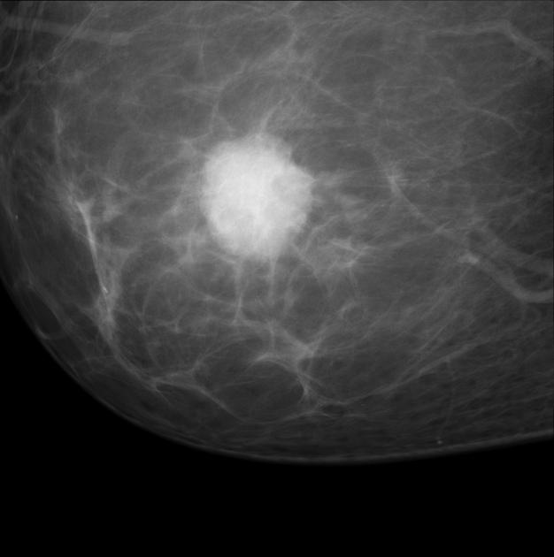
Radiographic appearance
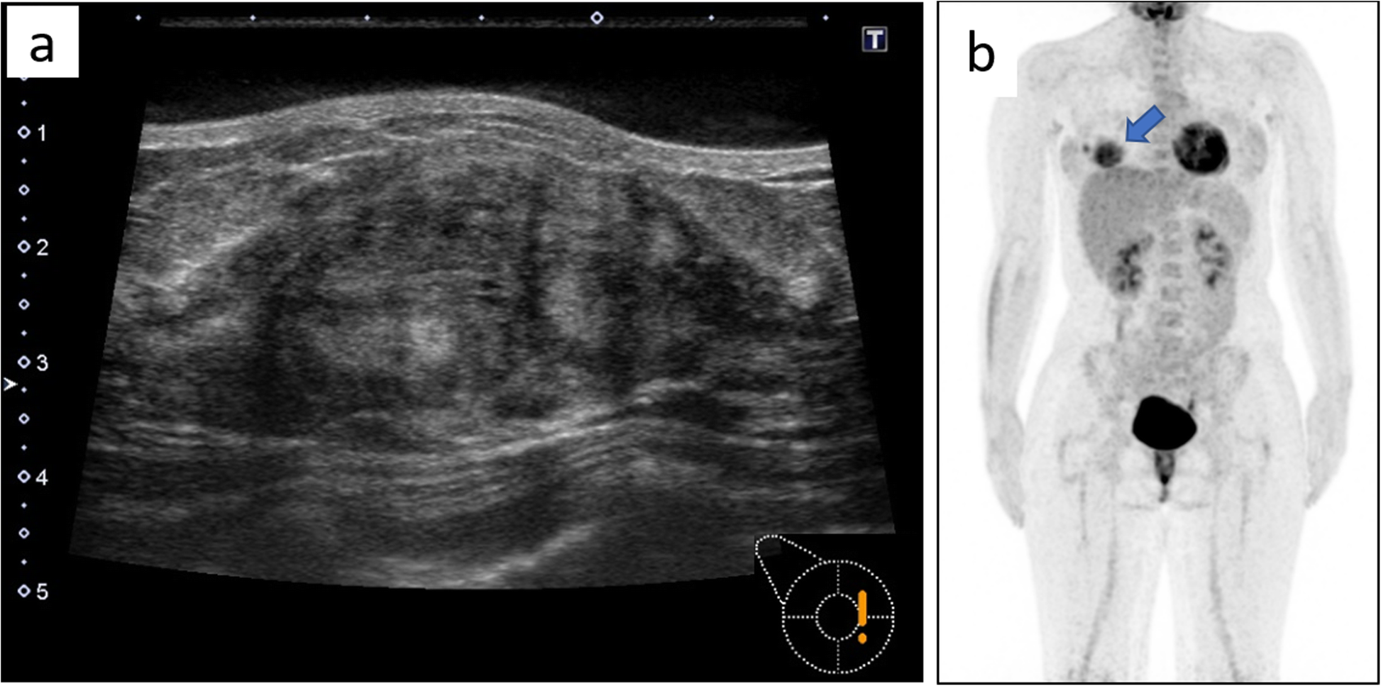
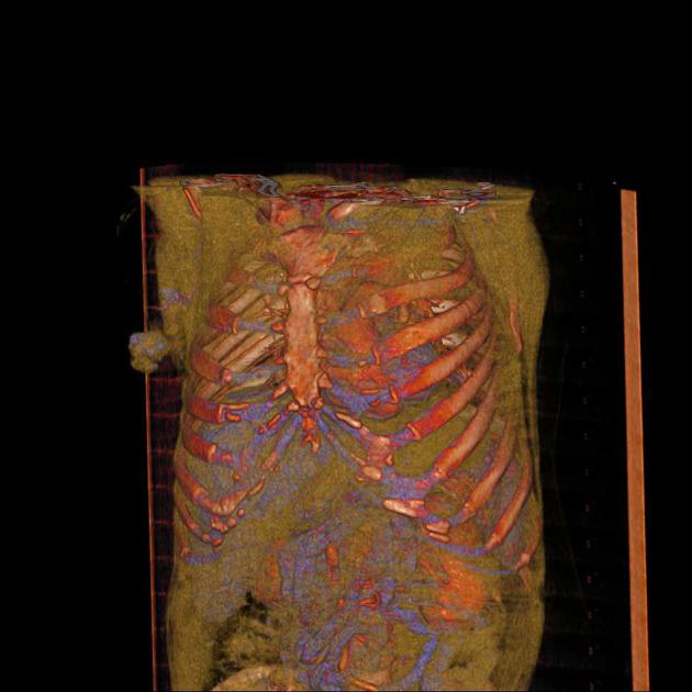
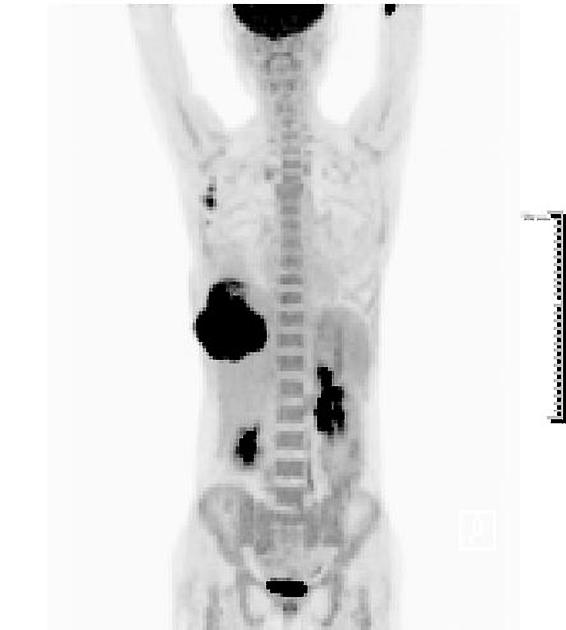
Dedicated evaluation of the breast involves multiple imaging modalities to detect and localize lesions for biopsy. In all modalities, regional metastasis can be suspected by the presence of axillary adenopathy.
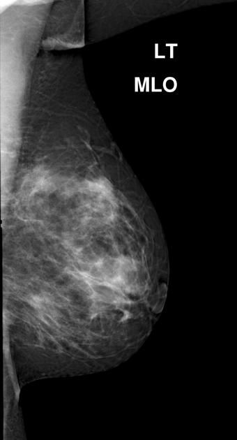
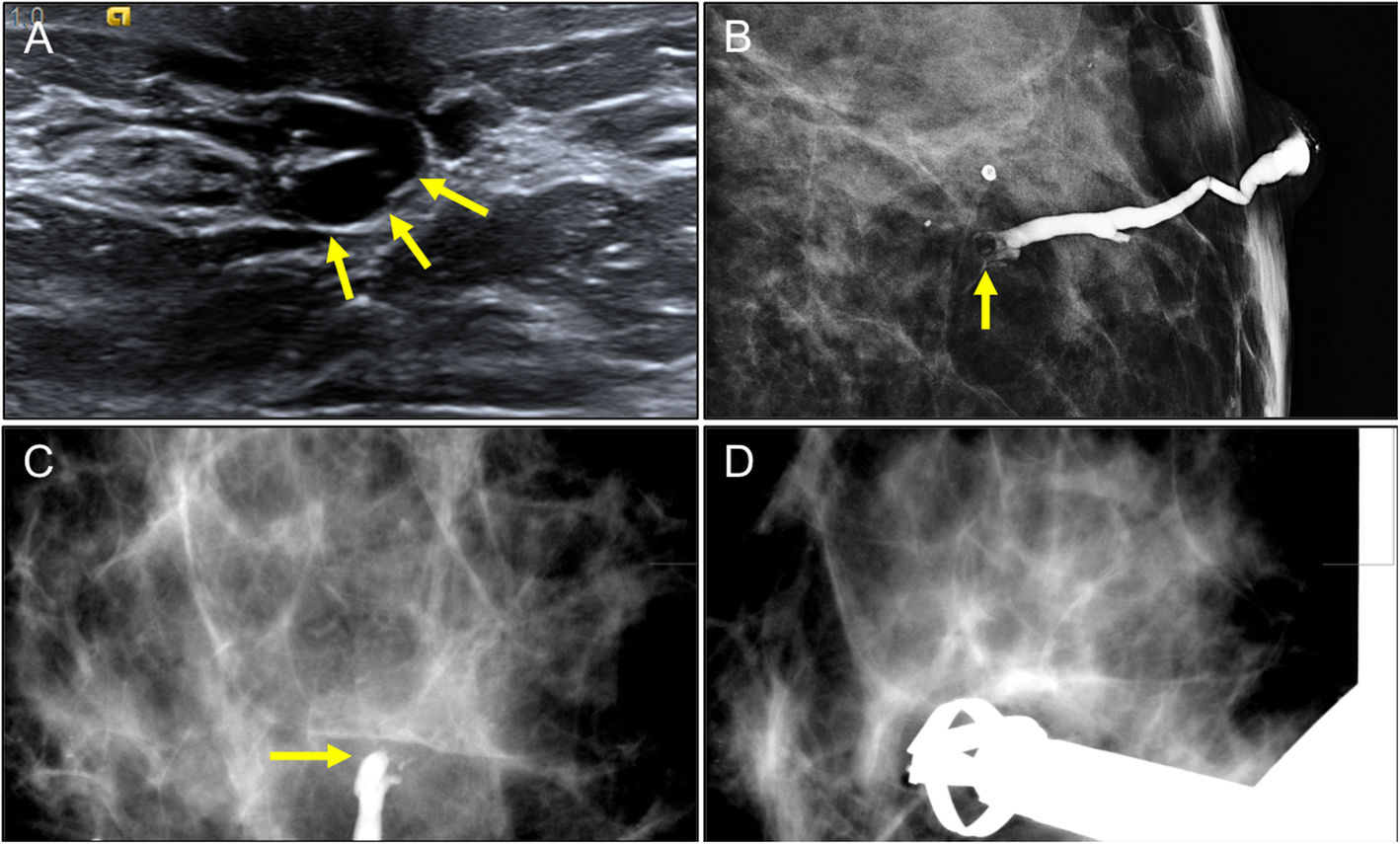
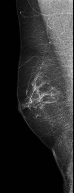
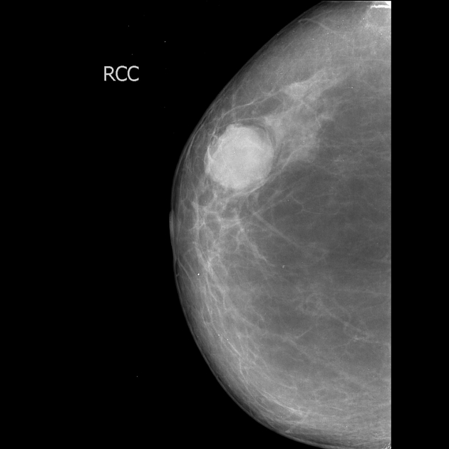
Mammography
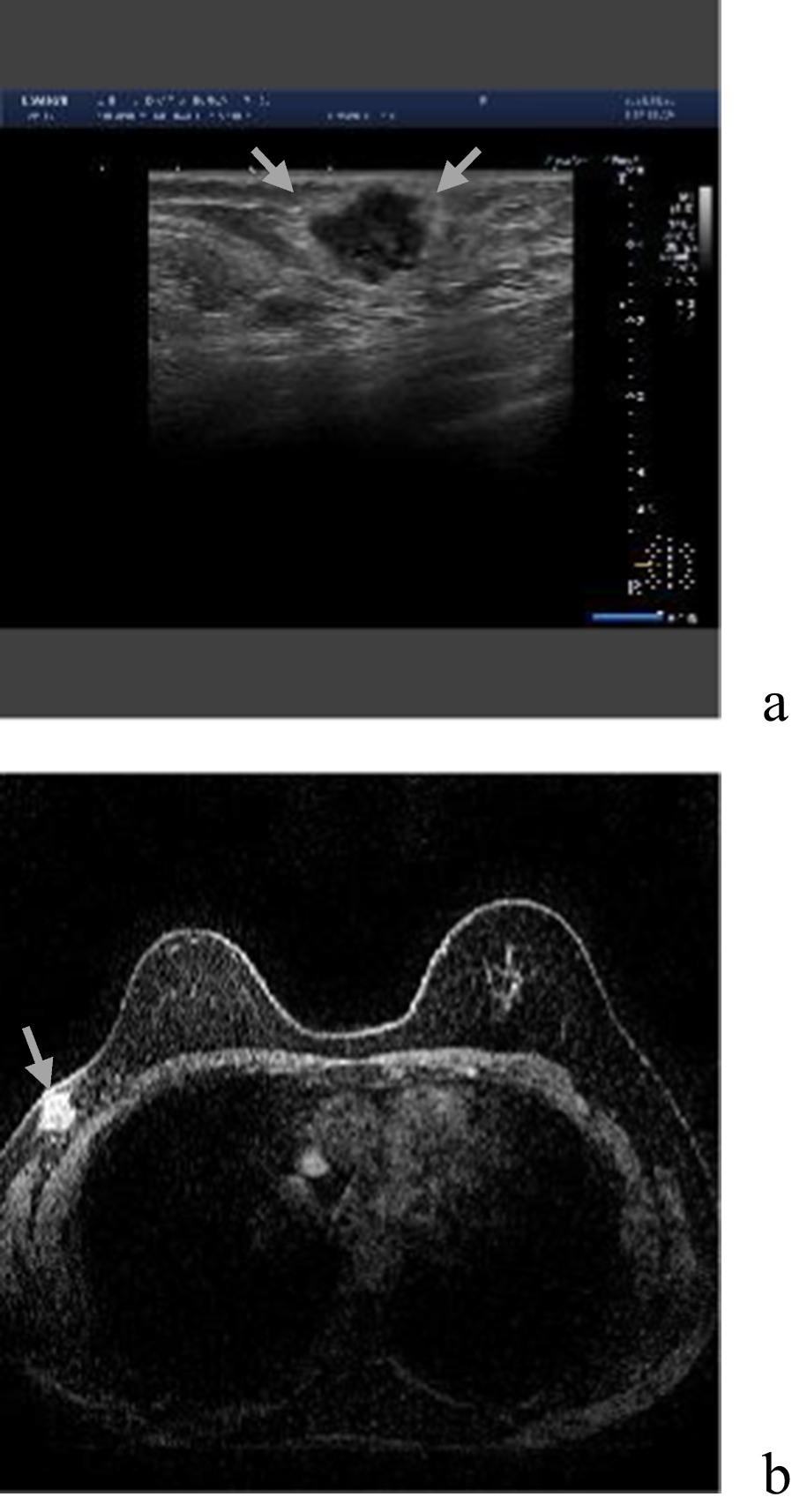
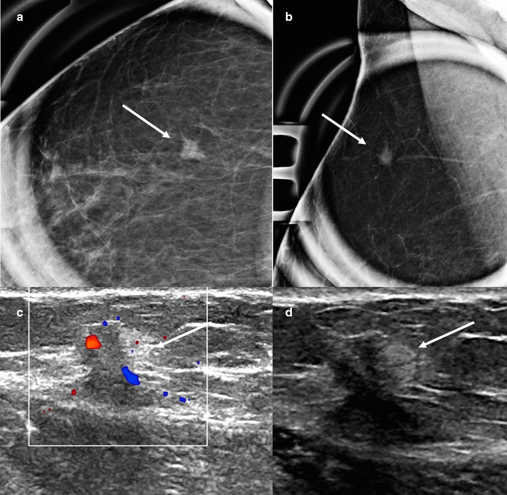
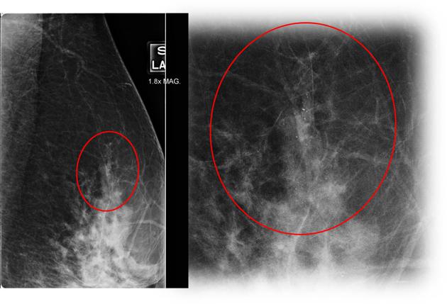
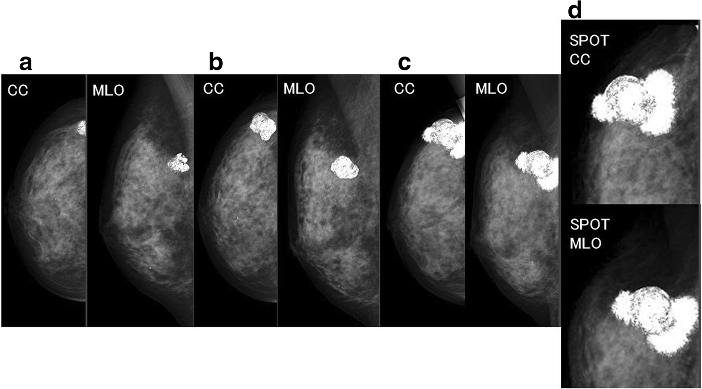
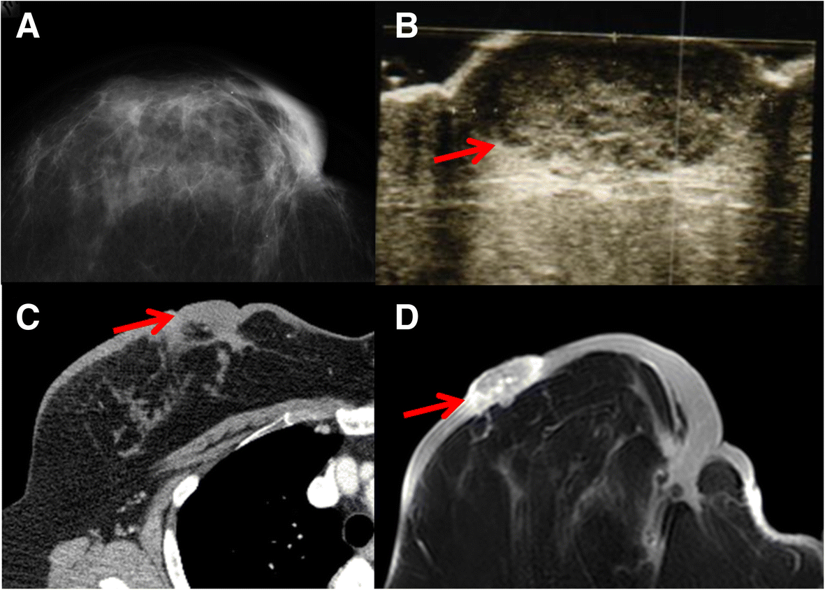
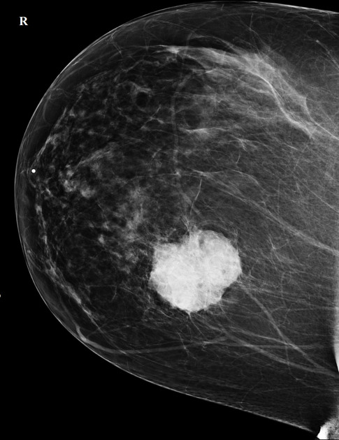
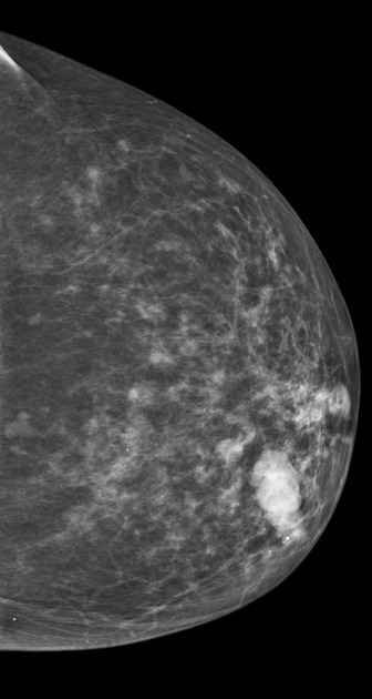
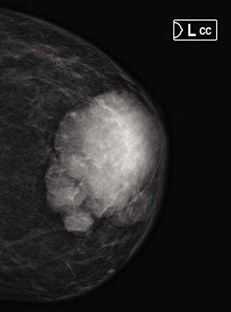
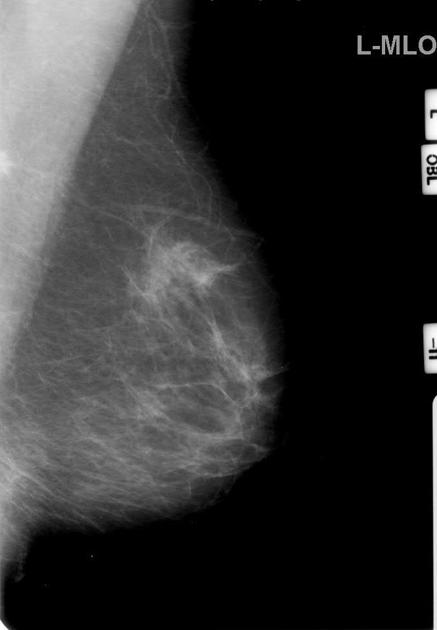
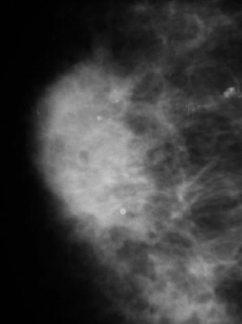
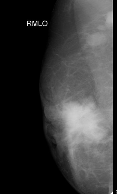
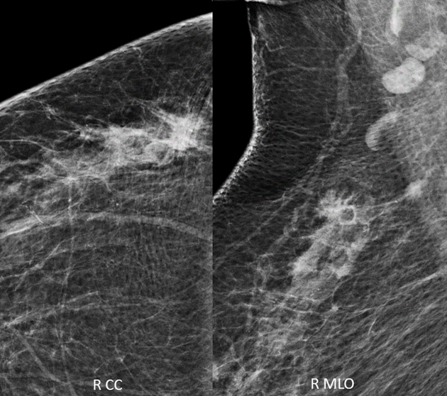
Neoplasms have varied appearances, including masses, asymmetries, calcifications, or architectural distortions.
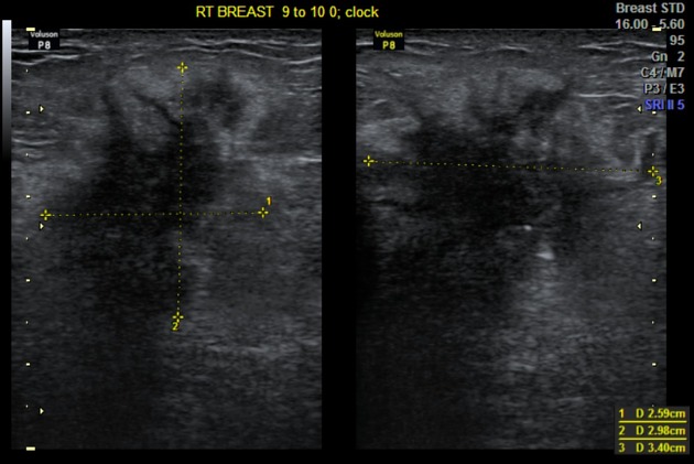
Ultrasound
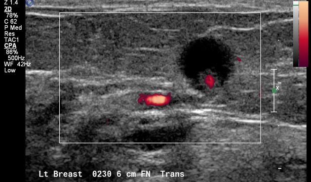
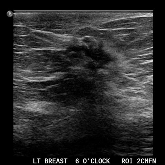
Neoplasms can appear as masses or architectural distortions. Calcifications can sometimes be seen.
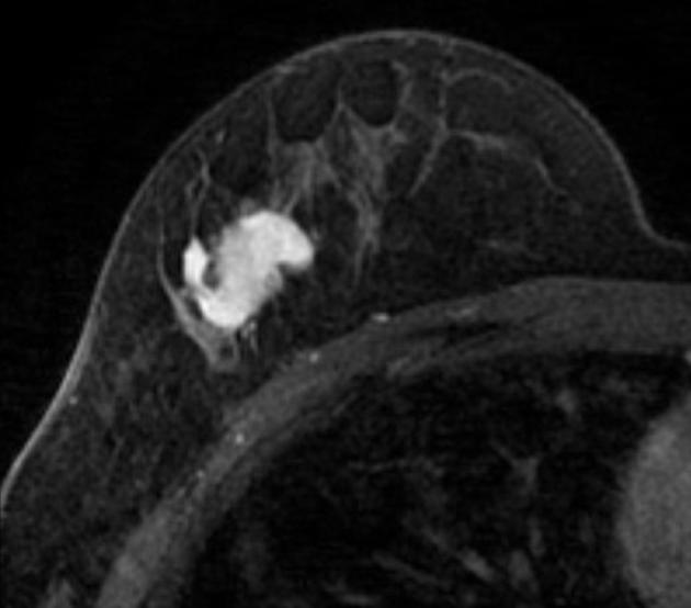
MRI
Neoplasms can manifest as masses with or without enhancement, nonmass enhancement, or foci of enhancement.
CT
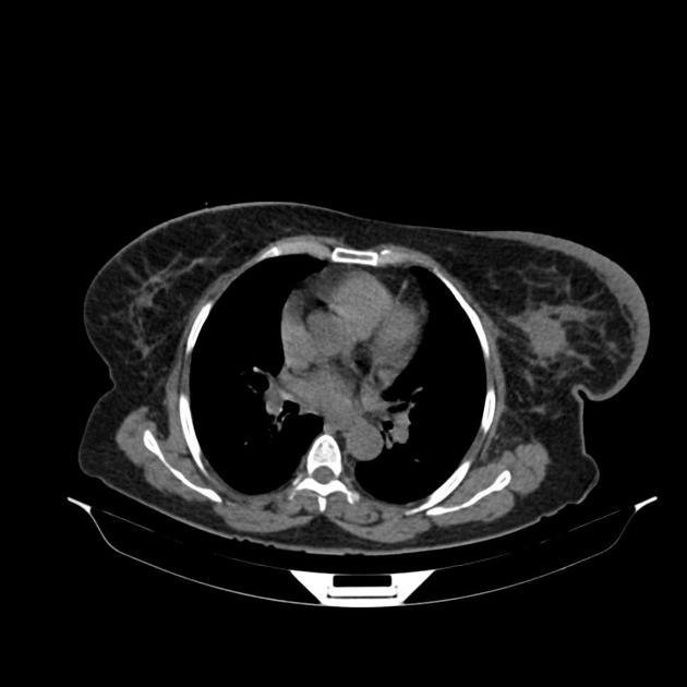
Breast masses may be incidentally identified but CT is not the preferred modality for dedicated breast evaluation. If calcifications are visualized on CT, they are nearly all benign .
Radiology report
The use of a standard lexicon is recommended to enhance communication with referrers and audit performance: breast imaging-reporting and data system (BI-RADS).
Siehe auch:
- invasives lobuläres Karzinom
- breast lumps
- Metastasen bei Mammakarzinom
- intraductales Papillom der Mamma
- Morbus Paget der Mamille
- Fibromatose der Mamma
- Sarkom der Mamma
- Mammakarzinom beim Mann
- artifacts that mimic breast calcification
- fibrosarcoma of the breast
- inflammatorisches Mammakarzinom
- Lymphom der Mamma
- Granularzelltumor der Mamma
- adenoid cystic carcinoma of the breast
- tubular carcinoma of breast
- lobular carcinoma in situ (LCIS)
- Liposarkom der Mamma
- apocrine carcinoma of the breast
- komplizierte Zyste Mamma
- duktales in situ Karzinom der Mamma
- Malignitätskriterien Sonographie Mamma
- tubulolobular carcinoma of breast
- extraskelettales Osteosarkom der Mamma
- Phylloidestumor
- medullary breast carcinoma
- Mammakarzinom Sonographie
- metaplastic carcinoma of the breast
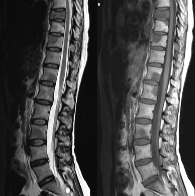
- metastasis to the breast
- comedo type
- Angiosarkom der Mamma
- breast screening
- Atypische duktale Hyperplasie (ADH)
- papilläres Mammakarzinom
- terminal duct lobular unit (TDLU)
- papilläre Neoplasien der Mamma
- metastasis(es) to breast
- invasives muzinöses Mammakarzinom
- atypische lobuläre Hyperplasie (ALH)
- Nicht-Komedo duktales in situ Karzinom der Mamma
- Juvenile Papillomatose der Mamma
- maligner Phylloidestumor
- Mammakarzinom Staging
und weiter:
- Tumoren der Schädelkalotte
- osteoblastische Knochenmetastasen
- solitäre lytische Läsion des Schädels
- Kerley-Linien
- cancer
- breast curriculum
- miliare Lungenherde
- tree in bud-Muster
- Metastasen in der Orbita
- bilaterale axilläre Lymphadenopathie
- Superscan Szintigraphie
- hyperdenser Lymphknoten
- breast ultrasound
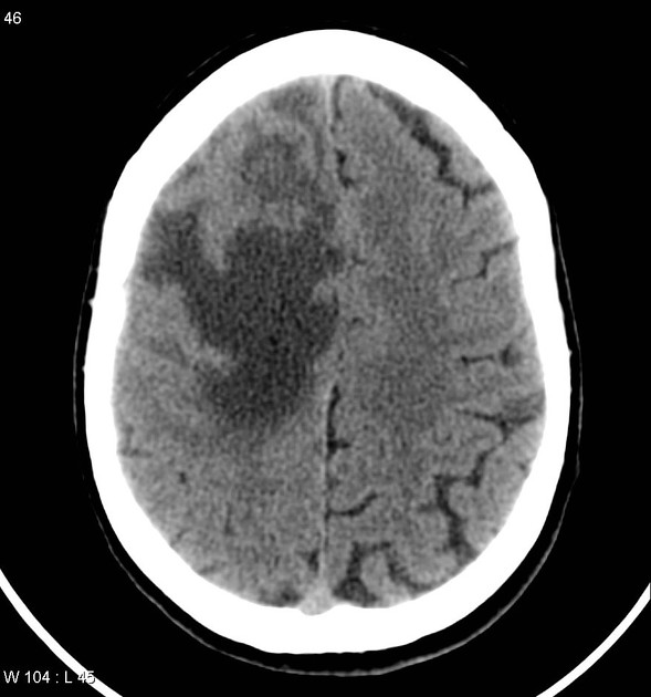
- metastases to the pituitary gland
- ultrasound appearances of liver metastases
- Mondor disease
- chronische abakterielle Mastitis
- Krukenberg-Tumor
- ivory vertebra sign
- differential diagnosis of unilateral axillary lymphadenopathy
- Somatostatin-Rezeptor-Szintigrafie
- miliary nodules in the exam
- diabetische Mastopathie
- tubular carcinoma of the breast
- FIGO-Klassifikation
- hyperechoic breast lesions
- einfache Zyste Mamma
- fibroadenomatoid mastopathy
- microglandular adenosis of the breast
- architectural distortion in mammography
- metastatic axillary lymphadenopathy of unknown primary
- asymmetrical density in mammography
- breast screening programmes
- metastases to the cervix
- Cowden-Syndrom
- differential diagnosis of calcific axillary lymphadenopathy
- LCIS
- Galaktozele
- medullary carcinoma of the breast
- Brachytherapie
- postoperative Narben Mamma
- triple receptor negative breast cancer
- lobular breast carcinoma
- ductal adenoma of breast
- pregnancy associated breast cancer
- intracystic papillary carcinoma of the breast
- verkalkte Metastasen
- differential diagnosis of dilated ducts on breast imaging
- multi-focal breast cancer
- bilateral lobular carcinoma of the breast
- breast self-examination
- differential diagnosis of dilated mammary veins
- multi-centric breast cancer
- Mammakarzinom in einer Zyste
- Brustdichte in der Mammographie
- Abszess der Mamma
- gemischt osteolytisch osteoblastische Knochenmetastasen
- idiopathische granulomatöse Mastitis
- Senologie
- granulomatöse Mastitiden allgemein
- scirrhous carcinoma of the breast
- eingeblutete Metastasen
- sklerosierende Adenose der Mamma
- radiäre Narbe der Mamma
- hyperdense pulmonale Raumforderungen
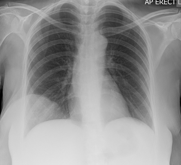
- Lungenmetastasen bei Mammakarzinom


 Assoziationen und Differentialdiagnosen zu Mammakarzinom: