Carpal bone fractures
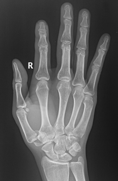
Trapezium
fracture • Trapezium fracture - Ganzer Fall bei Radiopaedia
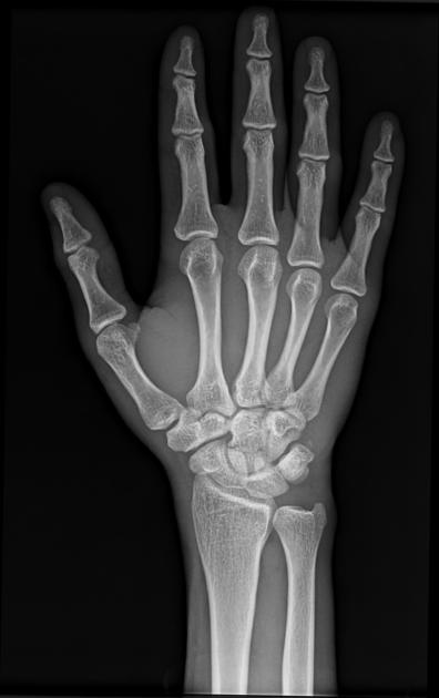
Scaphoid
fracture • Scaphoid fracture - Ganzer Fall bei Radiopaedia
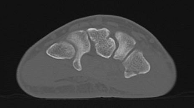
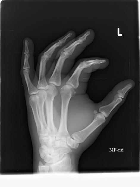
Pisiform
fracture • Pisiform fracture - Ganzer Fall bei Radiopaedia
Hook of
hamate fracture • Fracture base of the hook of the hamate - Ganzer Fall bei Radiopaedia
Capitate
fracture • Isolated capitate fracture - Ganzer Fall bei Radiopaedia
Trapezium
fracture • Trapezium fracture - Ganzer Fall bei Radiopaedia
Trapezoid
fracture • Isolated trapezoid fracture - Ganzer Fall bei Radiopaedia
Capitate
fracture • Isolated capitate fracture - Ganzer Fall bei Radiopaedia
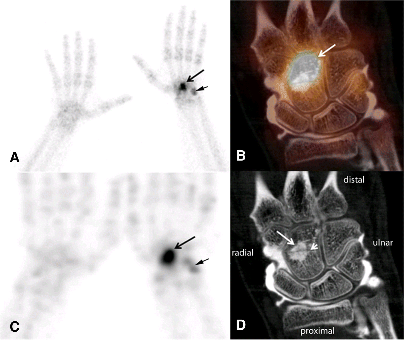
Occult
capitate fracture through a bone island – SPECT/CT arthrography imaging. SPECT/CT arthrography in a patient 1 month after carpal trauma with capitate fracture (short arrow in D) through a bone island (long arrow in D). Planar bone scintigraphy (a) SPECT (c) and SPECT/CT (b) with increased uptake in the capitate (arrow). SPECT/CT arthrography (b). CT arthrography (d) showing intact ligaments and cartilages. Additionally, slightly increased traumatic uptake in the pisiforme bone (short arrow) is visible on planar images and SPECT

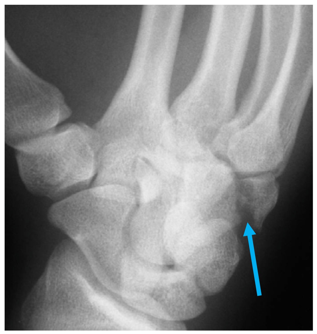
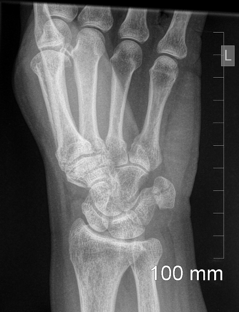
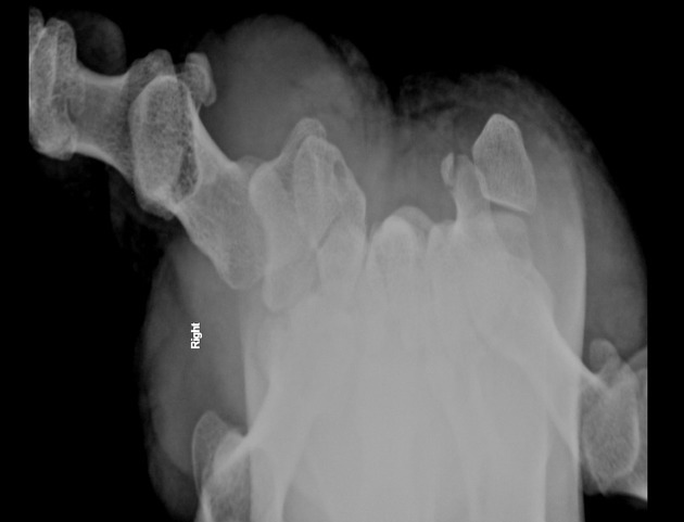
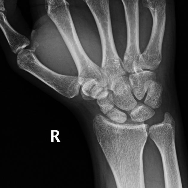
Lessons to be
learned from a missed case of Hamate fracture: a case report. Pronated oblique 30° x-ray view. Blue arrow shows fracture site.
Trapezium
fracture • Trapezium fracture - Ganzer Fall bei Radiopaedia
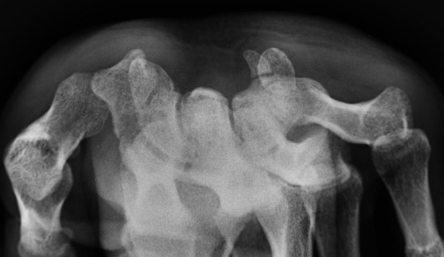
Hook of
hamate fracture • Scaphoid and hook of hamate fractures - Ganzer Fall bei Radiopaedia
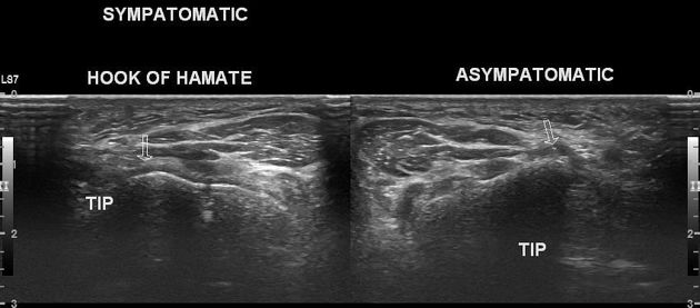
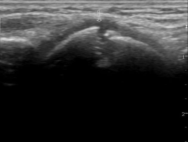
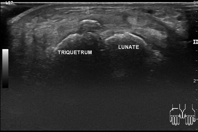
Hook of
hamate fracture • Hook of hamate fracture (ultrasound) - Ganzer Fall bei Radiopaedia
Hook of
hamate fracture • Hook of hamate fracture - Ganzer Fall bei Radiopaedia
Hook of
hamate fracture • Hook of hamate fracture - Ganzer Fall bei Radiopaedia
Hook of
hamate fracture • Hook of hamate fracture - Ganzer Fall bei Radiopaedia
Pisiform
fracture • Isolated pisiform fracture - Ganzer Fall bei Radiopaedia
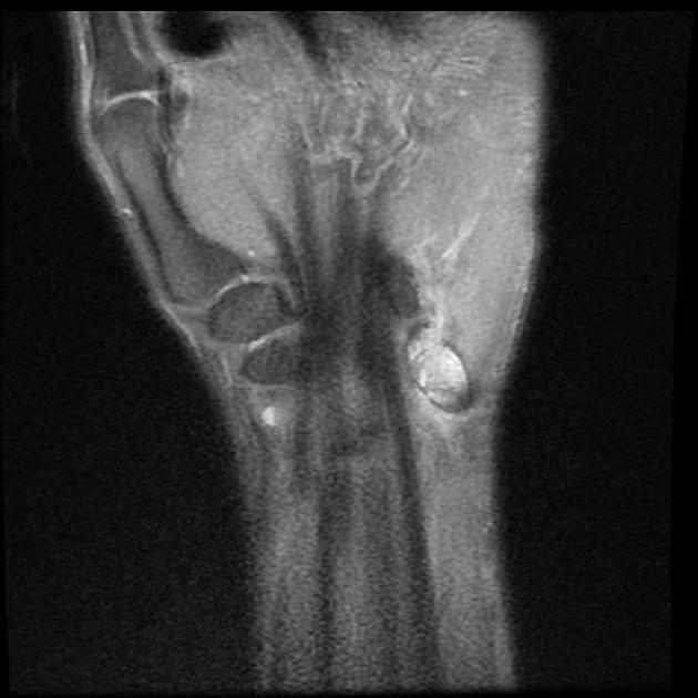
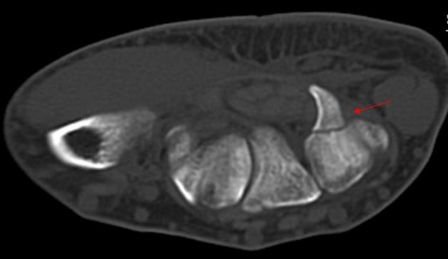
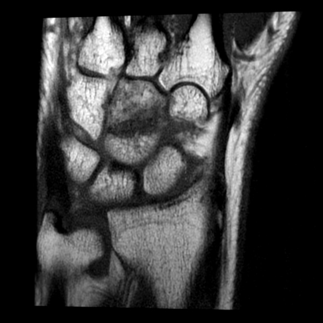
Pisiform
fracture • Isolated pisiform bone fracture (MRI) - Ganzer Fall bei Radiopaedia
Pisiform
fracture • Pisiform fracture - Ganzer Fall bei Radiopaedia


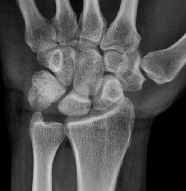
Triquetral
fracture • Triquetral fracture - Ganzer Fall bei Radiopaedia
Triquetral
fracture • Triquetral fracture - Ganzer Fall bei Radiopaedia
Triquetral
fracture • Triquetral fracture - Ganzer Fall bei Radiopaedia
Triquetral
fracture • Triquetral fracture - Ganzer Fall bei Radiopaedia
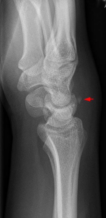
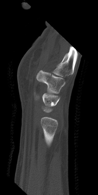
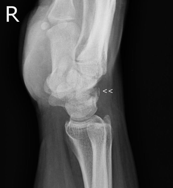
Typisches
seitliches Röntgenbild eines kleinen Ausbruchs aus dem Os triquetrum nach dorsal (Pfeil). Man erkennt auch die deutliche Schwellung des Weichteils über der Fraktur.
Lunate
fracture • Lunate fracture - Ganzer Fall bei Radiopaedia
Scaphoid
fracture • Scaphoid fracture - Ganzer Fall bei Radiopaedia

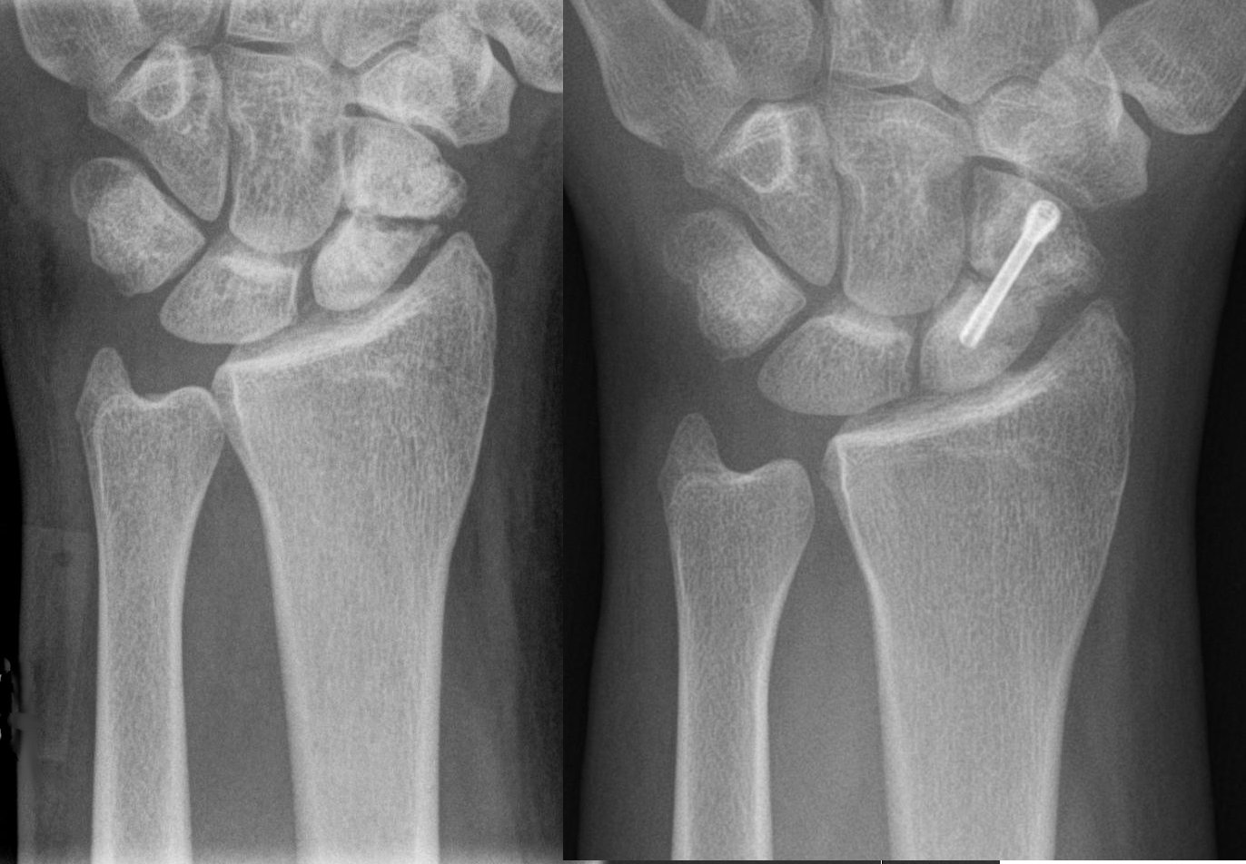
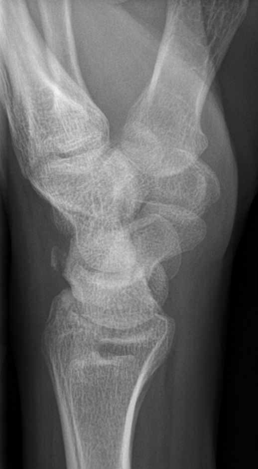
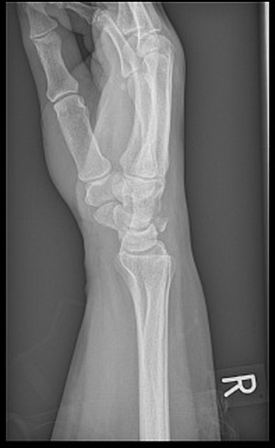
Scaphoid
fracture. Scaphoid fracture in 1/3 proximal of the scaphoid and neutral ulnar variance. There are no other fractures or signs of small parts lesion.
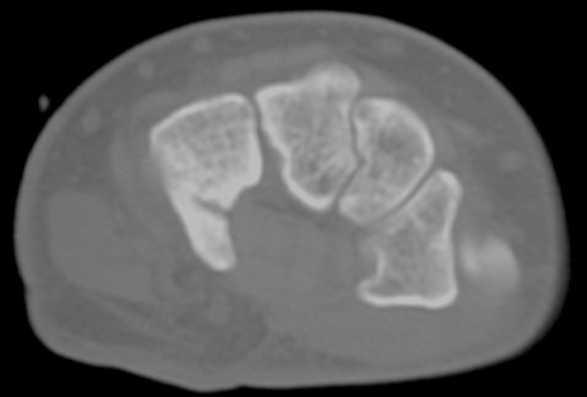
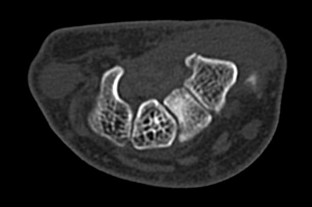
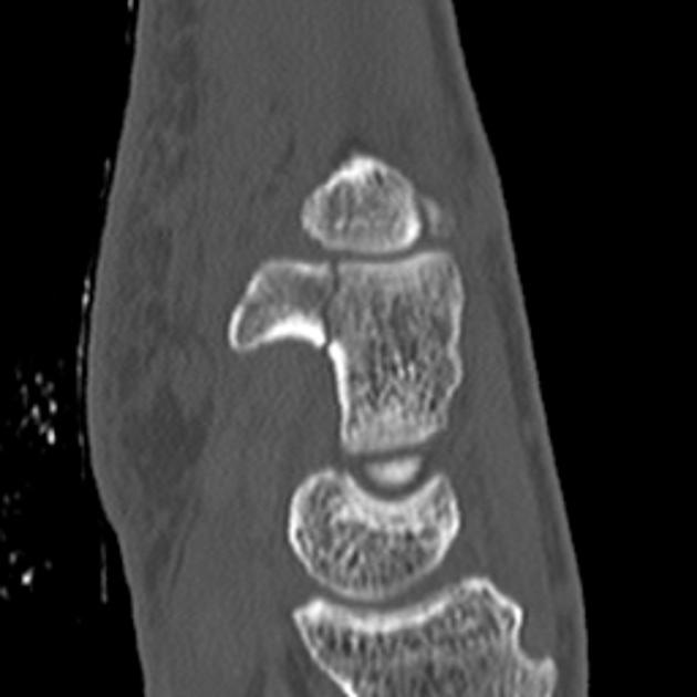
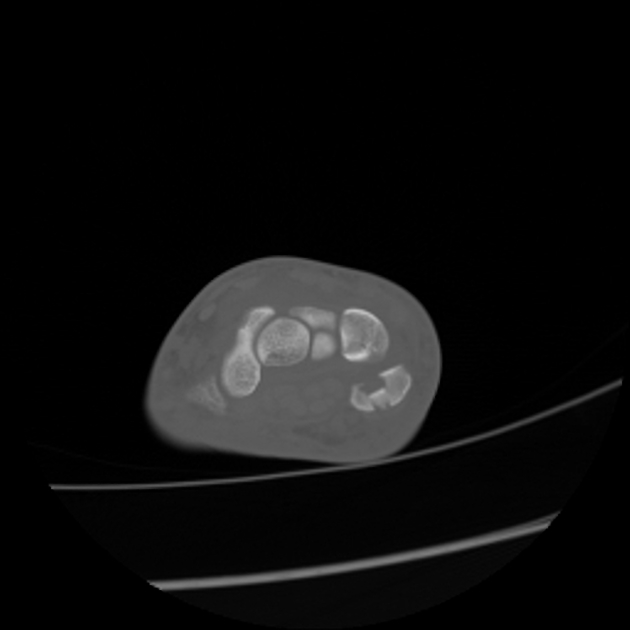
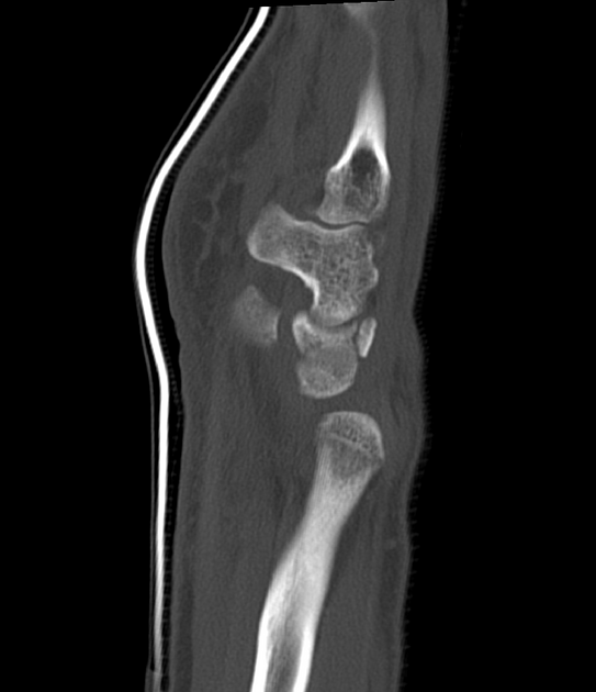
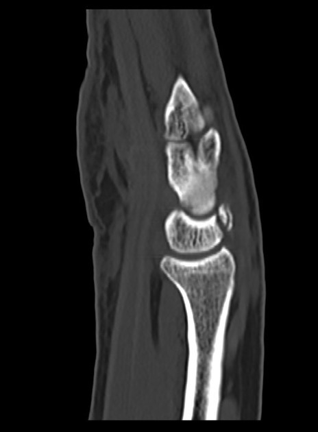
Case report
of right hamate hook fracture in a patient with previous fracture history of left hamate hook: is it hamate bipartite? . CT of left wrist indicating hamate hook fracture.
Carpal bone fractures comprise a range of different fractures which carry varying outcomes. They can involve one or a combination of carpal bones and also be part of fracture-dislocations.
Individual fractures include
- scaphoid fracture: 50-80%
- lunate fracture: 3.9%
- triquetral fracture: ~18%
- pisiform fracture: 1.3%
- trapezium fracture: ~3-5%
- trapezoid fracture: 0.4%
- capitate fracture: 1.9%
- hamate fracture: 1.7%
Fracture dislocations include
- perilunate dislocation: considered most common
- perilunate fracture-dislocation
- transradial styloid perilunate fracture-dislocation
- transcapitate perilunate fracture-dislocation
- transtriquetral perilunate fracture-dislocation
- transulnar styloid perilunate fracture-dislocation
- lunate dislocation
See also
Siehe auch:
- Triquetrumfraktur
- Fraktur Os pisiforme
- Os capitatum Fraktur
- Scaphoidfraktur
- Fraktur Os trapezium
- Fraktur Os hamatum
- Fraktur Os trapezoideum
- Frakturen Os lunatum
und weiter:


 Assoziationen und Differentialdiagnosen zu Frakturen der Handwurzelknochen: