gartner duct cyst

Gartner duct cysts develop from embryologic remnants of the Wolffian (mesonephric) duct. They are often noticed incidentally on ultrasound or MRI.
Clinical presentation
They may cause mass effect on adjacent structures.
Pathology
Location
Gartner duct cysts are located in the anterolateral wall of the proximal (superior) portion of the vagina and are typically located above the level of the most inferior aspect of the pubic symphysis.
Histology
Like other cysts, they are lined with non-mucinous cuboidal or columnar epithelium.
Associations
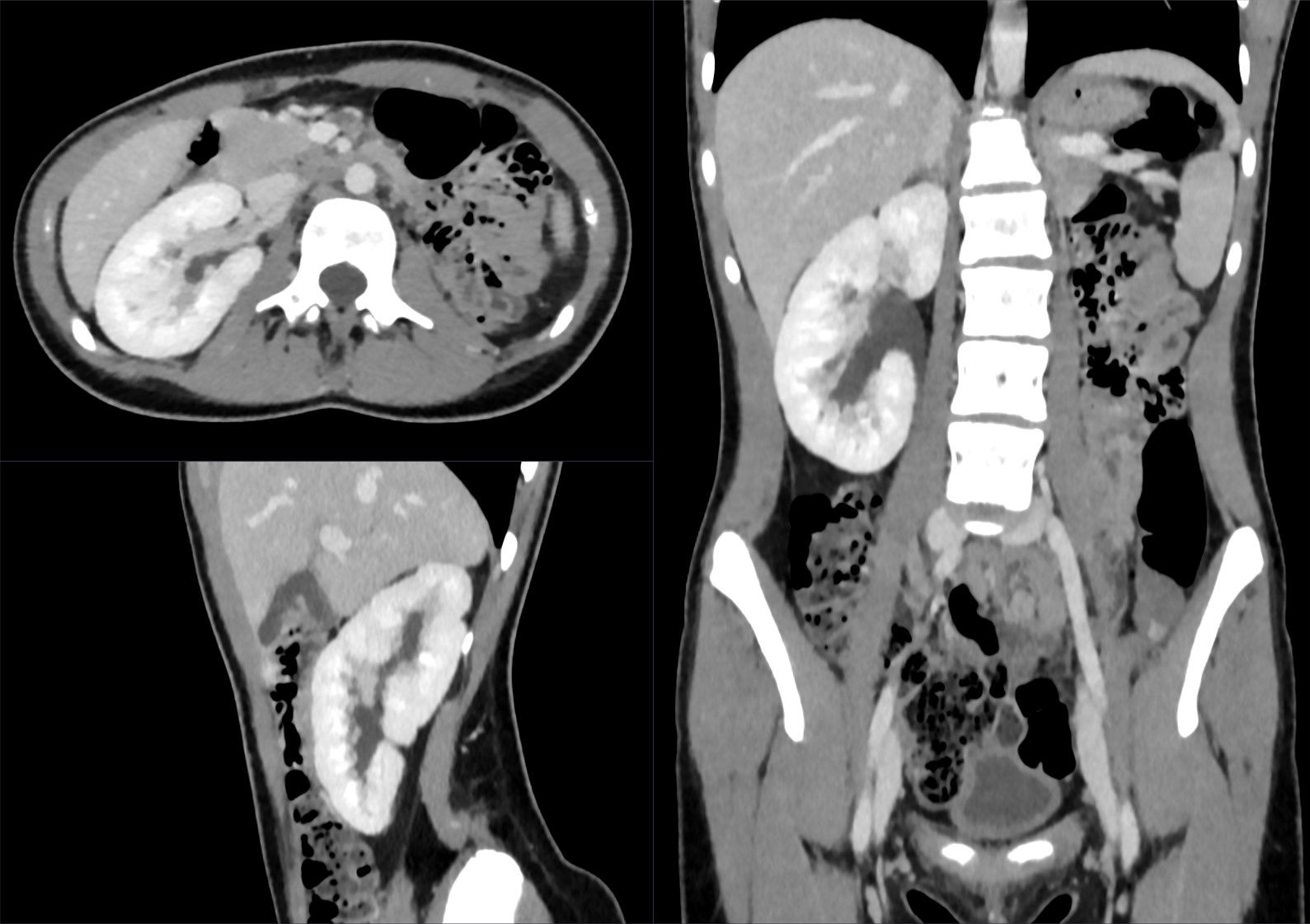
Gartner duct cysts most often are isolated findings, but can also be associated with abnormalities of the metanephric urinary system or in Herlyn-Werner-Wunderlich syndrome
- renal agenesis
- ipsilateral renal dysplasia
- cross fused ectopia
Radiographic features
Typically, they are simple cystic lesions arising from the anterolateral aspect of the superior vagina. They are usually small (<2 cm) although occasionally they can become very large (up to several centimeters) .
Complications
In rare cases with larger cysts, dyspareunia and problems in obstetric delivery have been described .
Differential diagnosis
General imaging differential considerations include:
- Bartholin gland cyst: their location at or below the level of the pubic symphysis and usually arising from posterolateral wall of the vagina; this helps to differentiate them from Gartner duct cysts
- urethral diverticulum: located around the urethra
Siehe auch:
- einseitige Nierenagenesie
- Bartholin-Zyste
- Divertikel der Urethra
- gekreuzte Nierendystopie mit Verschmelzung
- Wolffian (mesonephric) duct
und weiter:


 Assoziationen und Differentialdiagnosen zu gartner duct cyst: