Humerus


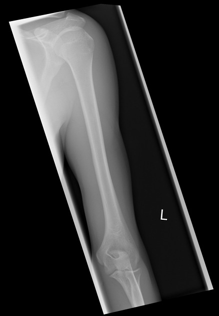
The humerus (plural: humeri) is a tubular bone of the arm that articulates proximally at the shoulder with the glenoid of the scapula, and distally at the elbow, with the radius and ulna.
Gross anatomy
Osteology
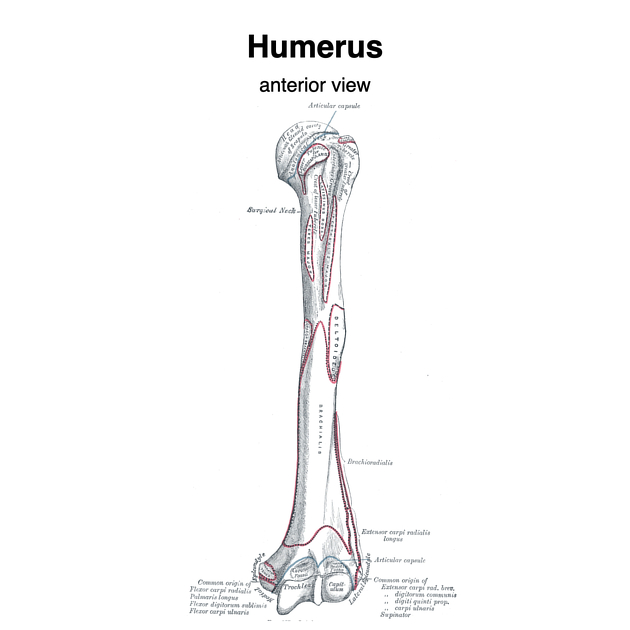
The humerus begins proximally as a rounded head and joins the greater and lesser tubercles via the anatomical neck of the humerus. The surgical neck is found just inferior to the tubercles where the shaft begins. The surgical neck is a common site for fractures (hence its name), while fractures of the anatomical neck are rare.
The shaft of the humerus has a cylindrical shape proximally then flattens to produce more distinct surfaces. There are three surfaces (anterolateral, anteromedial and posterior surfaces) and three borders (anterior, lateral and medial borders).
The condyle of the humerus articulates with the radius and ulna. It is composed of the trochlea, capitulum, and medial and lateral epicondyles.
The supracondylar process is a variant that is found ~5 cm above the medial epicondyle and can be 2 to 22 mm in length. It can be continuous with the ligament of Struthers, beneath which the median nerve and brachial artery pass.
The proximal and distal ends of the humerus are cancellous bone with a superficial layer of compact bone. The shaft is comprised of compact bone containing a medullary canal traversing its length.
Articulations
- proximal (refer to shoulder joint article for more details):
- head of the humerus: articulates with the glenoid cavity of the scapula
- distal (refer to elbow joint article for more detail):
- the condyle of the humerus:
- trochlea: articulates with the trochlear notch of the ulna
- radial fossa: receives the anterior border of the radial head in forearm flexion
- capitulum: articulates with the head of the radius
- olecranon fossa: receives the olecranon process of the ulna with forearm extension
- coronoid fossa: receives the coronoid process of the ulna with forearm flexion
- the condyle of the humerus:
Attachments
- greater tubercle: insertion of the supraspinatus superiorly, infraspinatus intermediately, and the teres minor inferiorly
- lesser tubercle: insertion of the subscapularis
- bicipital groove: located between the greater and lesser tubercles, is the insertion of the latissimus dorsi and contains the long head of the biceps brachii. It is enclosed by the small broad transverse humeral ligament.
- deltoid tuberosity: insertion of the deltoid
- anterior-medial surface: insertion of the latissimus dorsi superiorly, partial insertion of the coracobrachialis intermediately, and origin of the brachialis inferiorly
- lateral border: partial insertion site of the teres minor and partial origin to the triceps brachii
- medial border: insertion of the teres major and coracobrachialis
- lateral supracondylar ridge: origin of the brachioradialis, extensor carpi radialis longus, triceps brachii, and attachment of the lateral intermuscular septum
- medial supracondylar ridge: origin of the brachialis, pronator teres, triceps brachii, and attachment of the medial intermuscular septum
- lateral epicondyle: origin of the supinator and some extensor muscles of the forearm, also attachment to the radial collateral ligament of the elbow
- medial epicondyle: origin of some flexor muscles of the forearm, pronator teres and attachment to the medial ulna collateral ligament of the elbow
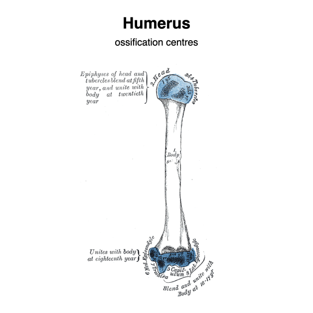
Development
There are eight ossification centers that begin ossification in the following order:
body (8 week of fetal life), head (1 year of age), capitulum (1 year), greater tubercle (3 years), lesser tubercle (5 years), medial epicondyle (5 years), trochlea (10 years), and lateral epicondyle (10 years).
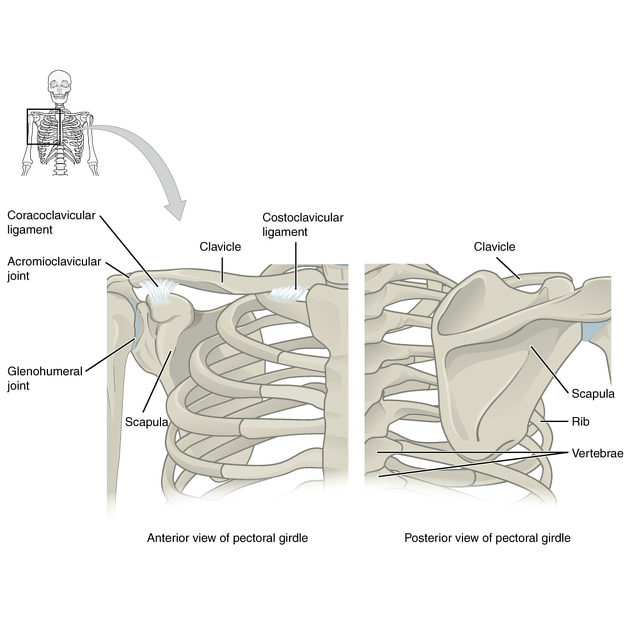
See also: ossification centers of the pectoral girdle; elbow ossification.
Anatomical variants
Related pathology
It may be fractured by excessive torsion or by a direct blow. The humeral neck is frequently fractured in elderly patients following a fall on the shoulder.
The humeral head may be dislocated from the glenoid fossa anteriorly or, much less frequently, posteriorly or inferiorly.
The humeral neck is also a common location for simple bone cysts.
See also
- proximal humeral fracture
- humeral shaft fracture
- lightbulb sign of posterior shoulder dislocation
- Hill-Sachs lesion
- Reverse Hill-Sachs lesion
- pseudocyst of the humerus
Siehe auch:
und weiter:


 Assoziationen und Differentialdiagnosen zu Humerus:

