Intravenous urography


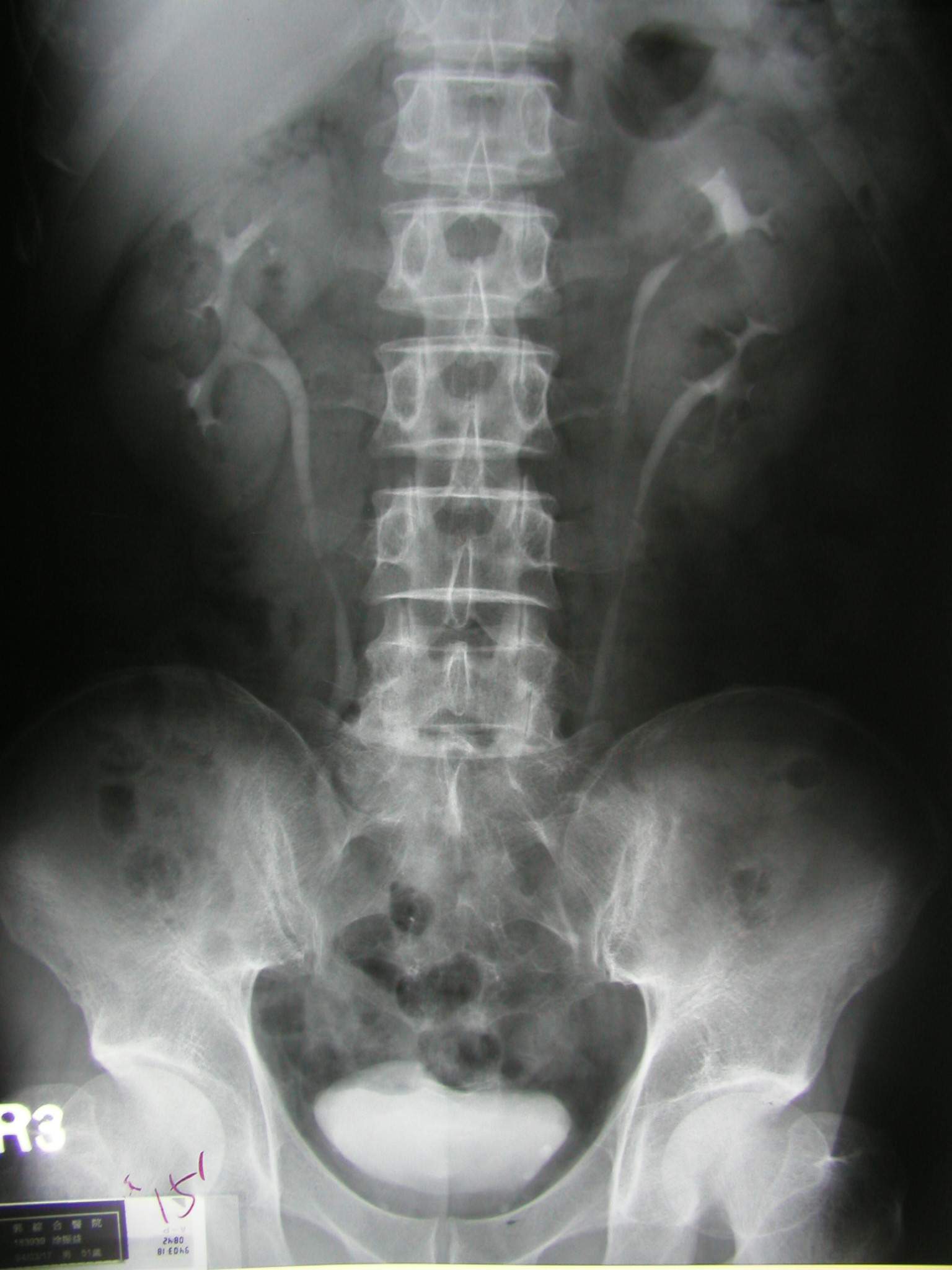

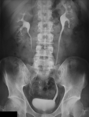
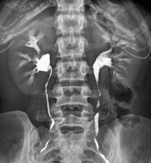
Intravenous urography (IVU), also referred to as intravenous pyelography (IVP) or excretory urography (EU), is a radiographic study of the renal parenchyma, pelvicalyceal system, ureters and the urinary bladder. This exam has been largely replaced by CT urography.
Terminology
Some prefer the term "urogram" to refer to visualization of the kidney parenchyma, calyces, and pelvis after intravenous injection of contrast, and reserve the term "pyelogram" to retrograde studies involving the collecting system. In practice, both terms are often used interchangeably.
Procedure
Indications
- check for normal function of kidneys
- check for anatomical variants or congenital anomalies (e.g. horse-shoe kidney)
- check the course of the ureters
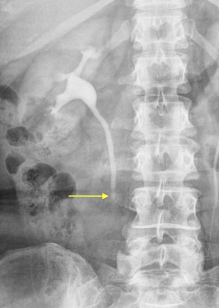
- detect and localize a ureteric obstruction (urolithiasis)
- assess for synchronous upper tract disease in those with bladder transitional cell carcinoma (TCC)
Patient preparation
- overnight fasting prior to the date of examination; a laxative would help to achieve a good preparation
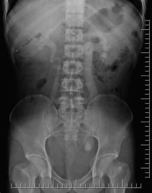
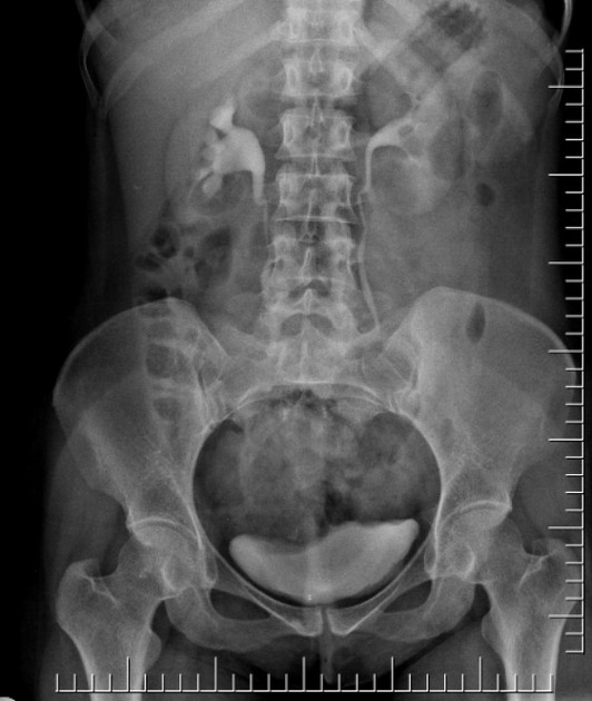
- on the day of the procedure take a scout/pilot film to check patient preparation and also for radiopaque calculi
- check serum creatinine level to be within the normal range (as per hospital guidelines)
- take a history of the patient for any known drug allergies followed by written informed consent for the procedure
Technique
Exposures are generally in the 65-75 kV range, mA of 600-1000, with exposure of <0.1 sec. Higher kV ranges reduce contrast of the renal parenchyma.
- IV access is required for administration of a water-soluble contrast
- nonionic contrast is preferred
- dose will vary as per the weight of the patient; generally up to 1.5 ml/kg body weight is well tolerated by patient
- the contrast dose is usually instilled at a fast (bolus) rate
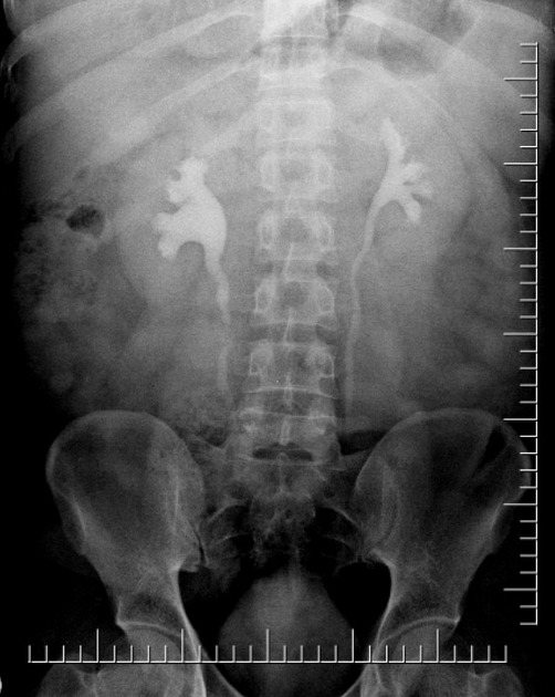
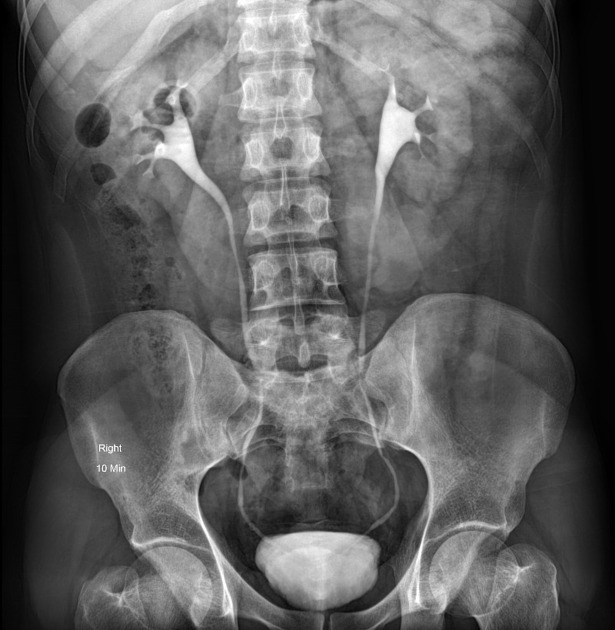
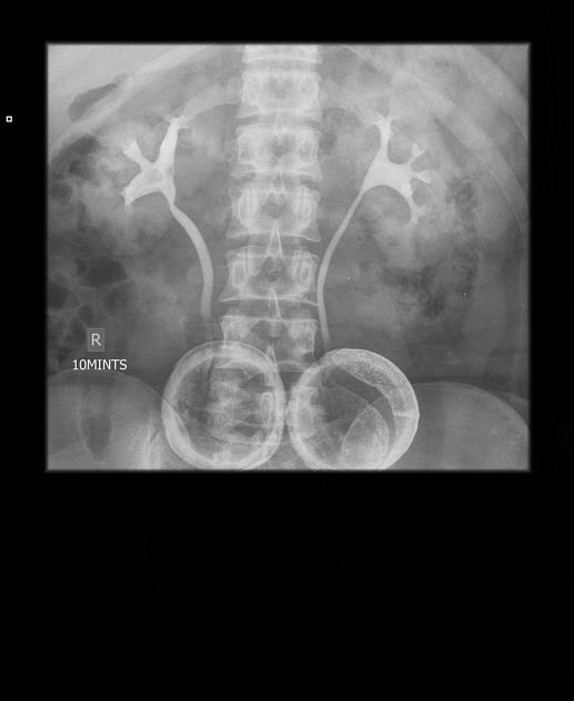
- the calyces are usually visualized in <2 minutes following contrast administration - this is the nephrogram
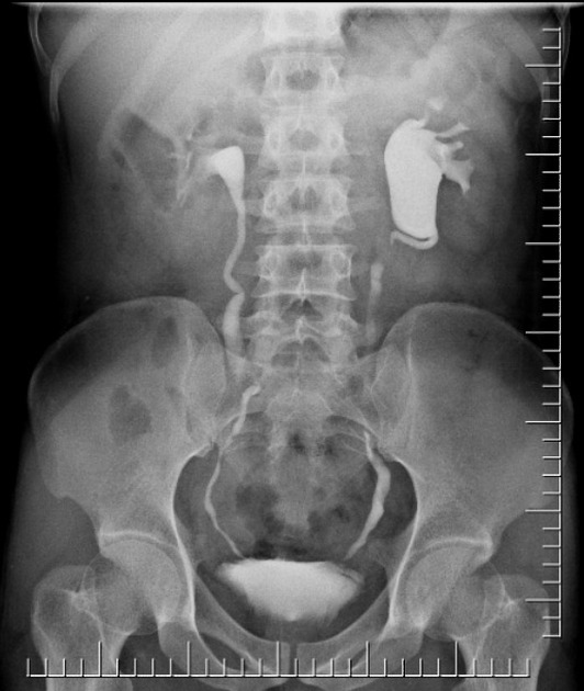
- serial images are taken at 5-20 minutes for visualization of the pelvicalyceal systems and ureters when required and with operator preference
- additional views taken are prone and obliques for ureters
- the full length 10-15 minute film is performed with a compression band applied to the patient
- compression should not be applied if ureteral calculi, ureteral obstruction, recent surgery, nephrostomy, or abdominal aortic aneurysm is suspected
- lastly take a full bladder and post-void film
There is a wide variation in protocols. One protocol is suggested below, but additional images should usually be obtained to answer the clinical question:
- scout images
- nephrogram (1-2 minutes)
- early and late images of the upper collecting system (abdominal compression then applied) (>3 minutes)
- tomography may be obtained, if desired
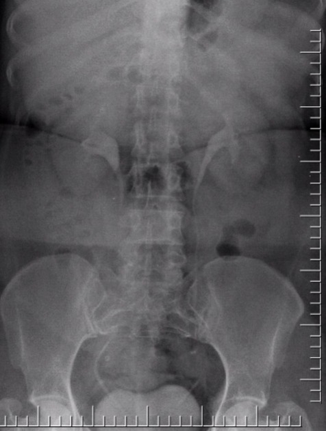
- supine, after release of compression, images of the upper collecting system and proximal ureters (10-15 minutes)
- supine image (20 minutes)
- prone image (20 minutes)
Emergency medications and emergency equipment must always be available in case the patient has a reaction to contrast.
Siehe auch:
und weiter:


 Assoziationen und Differentialdiagnosen zu Urografie: