Konzentrische Sklerose Baló
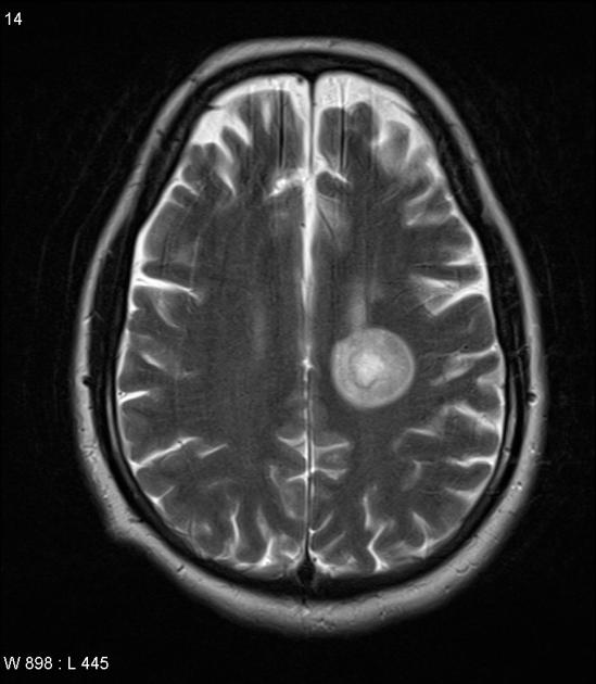
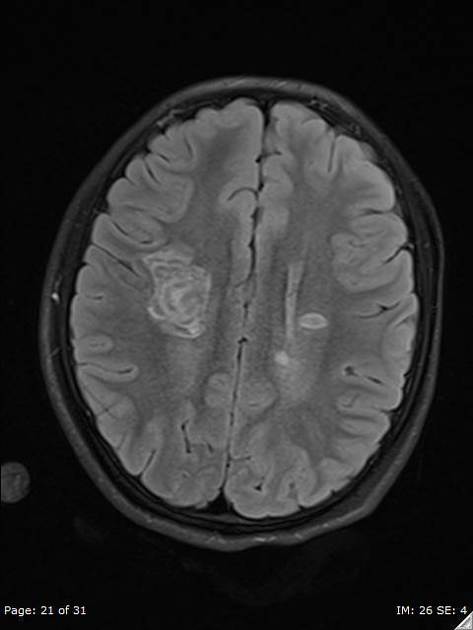
Baló concentric sclerosis is a rare and severe monophasic demyelinating disease, considered a subtype of multiple sclerosis, appearing as a rounded lesion with alternating layers of high and low signal intensity on MRI, giving it a characteristic 'bullseye' or 'onion bulb' appearance .
Epidemiology
It is a rare disorder most commonly found among people of Han Chinese and Filipino descent, although multiple sclerosis in these populations is less common than in Caucasians .
Clinical presentation
The presentation can be similar to other forms of multiple sclerosis, although it is usually monophasic, most closely resembling acute Marburg type multiple sclerosis, with rapid progression and sometimes fulminant course .
Pathology
Concentric layers of alternating demyelination and preserved myelin are seen, resulting in the characteristic imaging appearance, although the exact mechanism remains unclear .
Radiographic features
MRI
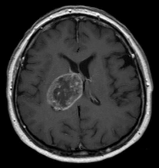
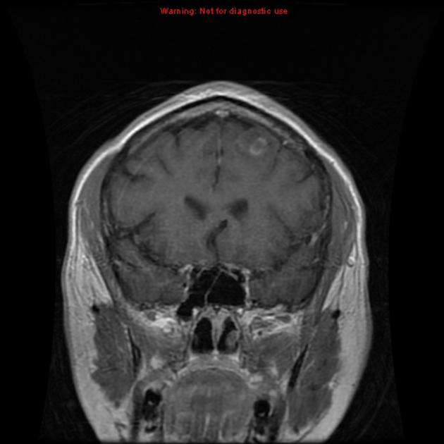
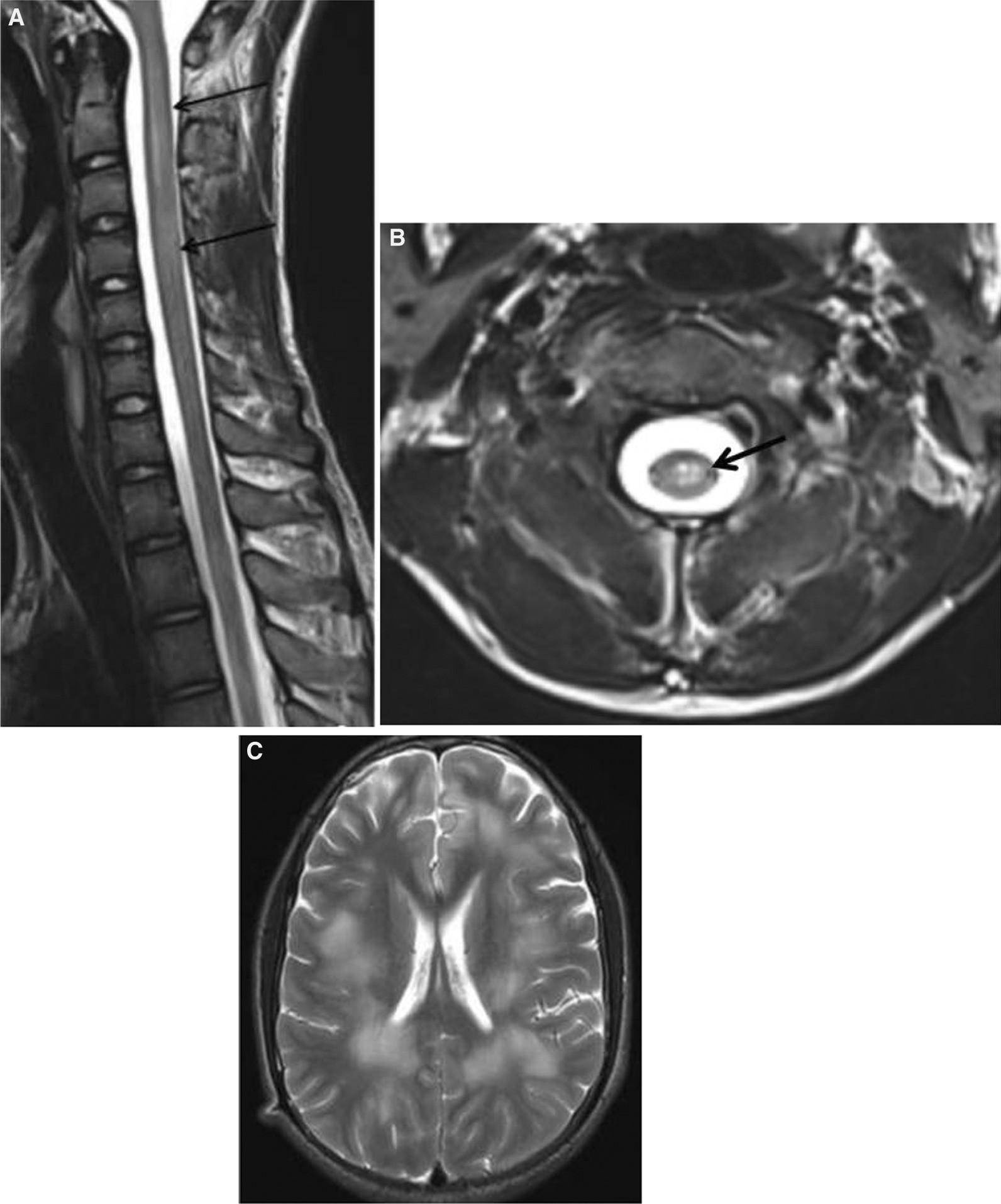
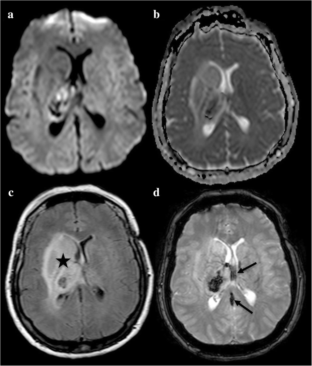
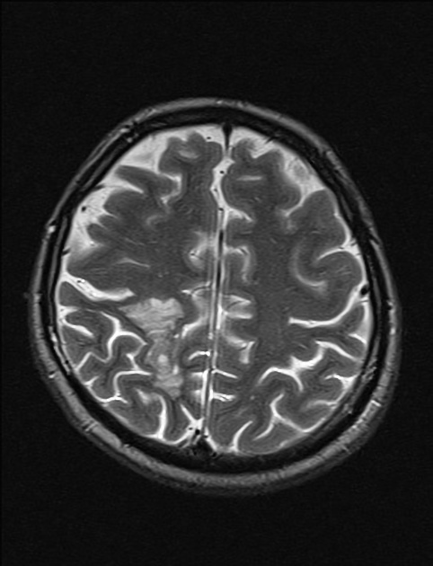
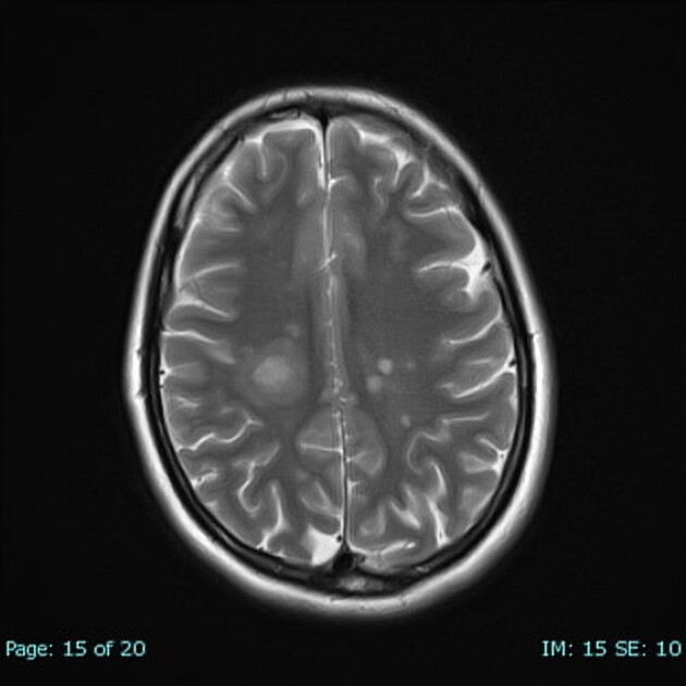
The characteristic feature is the development of alternating bands of demyelinated and myelinated white matter, which is seen as concentric rings or irregular stripes of high or low signal depending on the sequence :
- T1: irregular concentric areas of iso and low signal
- T2: irregular concentric areas alternating iso/hypointense and hyperintense signal
- T1 C+ (Gd): typically a peripheral ring of enhancement in the area of active demyelination
- DWI: some restricted diffusion in the outer ring
Treatment and prognosis
As with other forms of multiple sclerosis, corticosteroid has been used. The condition tends to have a rapidly progressive course but not necessarily fatal .
History and etymology
It is named after József Baló (1895–1979), Hungarian neuropathologist who first published the unusual case and subsequent death of a young law student who presented with fulminantly deteriorating, focal neurological symptoms in 1928 . "Leuko-encephalitis periaxialis concentrica" was the term he proposed to be used to the concentric focus of gray and white layers in the white matter he found during autopsy .
Differential diagnosis
Imaging differential considerations include:
- Marburg variant of MS and tumefactive demyelination
- both lack concentric pattern
- ADEM
- concentric ring pattern not present
- the patient usually has a preceding viral infection
- CNS lymphoma
- concentric ring pattern not present
- may have more mass effect
- CNS tumors (high-grade glioma or cerebral metastasis)
- cerebral abscess
- although abscesses often have a low T2 signal rim separating the fluid core and the surrounding enhancing rim, they do not possess alternating rings
- cerebral toxoplasmosis
Siehe auch:
- Hirnmetastase
- Glioblastoma multiforme
- Tumefaktive Multiple Sklerose
- Akute disseminierte Enzephalomyelitis
- zerebrale Venenthrombose
und weiter:


 Assoziationen und Differentialdiagnosen zu Balo concentric sclerosis: