Lipofibromatosis
Lipofibromatosis refers to a rare unspecified/borderline slow-growing soft tissue tumor prone to recurrence, which is often found in the hands and feet of children.
Terminology
Infantile or juvenile fibromatosis variant is an alternative term, which has been used but is now discouraged .
Epidemiology
Lipofibromatosis is typically found in childhood. Approximately 20% of the cases are detected at birth and roughly 50% are diagnosed in the first year of life. There is apparently a predilection for the male gender .
Associations
Lipofibromatosis has been associated with macrodactyly of the foot .
Clinical presentation
The typical presentation of lipofibromatosis is a slowly growing subcutaneous mass .
Pathology
Lipofibromatosis is an infiltrative soft-tissue tumor characterized by mature fatty tissue, fibrous fascicles made up of bland spindle cells and interposed fibroblasts, found between the first two tissue components .
Etiology
The etiology of lipofibromatosis is unknown .
Location
The most common location of lipofibromatosis is the subcutaneous tissues of hand and feet, though it can also involve skeletal muscle or occur at locations as the trunk or the head and neck region .
Macroscopic appearance
Macroscopically lipofibromatosis is characterized as poorly circumscribed tumors of yellow-white appearance and poorly discernable margins and firm consistency .
Microscopic appearance
Microscopic features of lipofibromatosis include the following :
- lobular architecture of mature fatty tissue cut across by fascicles of bland spindle cells within a collagenous matrix
- interspersed univacuolated cells at the interface of adipose tissue and spindle cell elements
- no or rare mitotic activity and nuclear atypia
- entrapment of adipose tissue or skeletal muscle fibers
Immunohistochemistry
Immunohistochemistry stains usually express CD34, smooth muscle actin and CD99 and might be positive for S100 or vimentin. They are usually negative for desmin, keratin and β-catenin .
Radiographic features
Ultrasound
Ultrasound can be used as a screening tool for the assessment of lipofibromatosis and might reveal a poorly circumscribed soft tissue mass with an intermittent hyperechoic signal intensity corresponding to the lipomatous components .
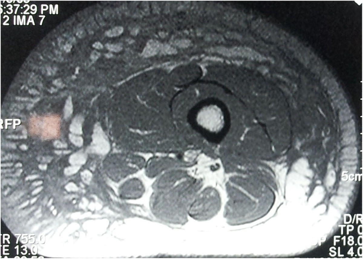
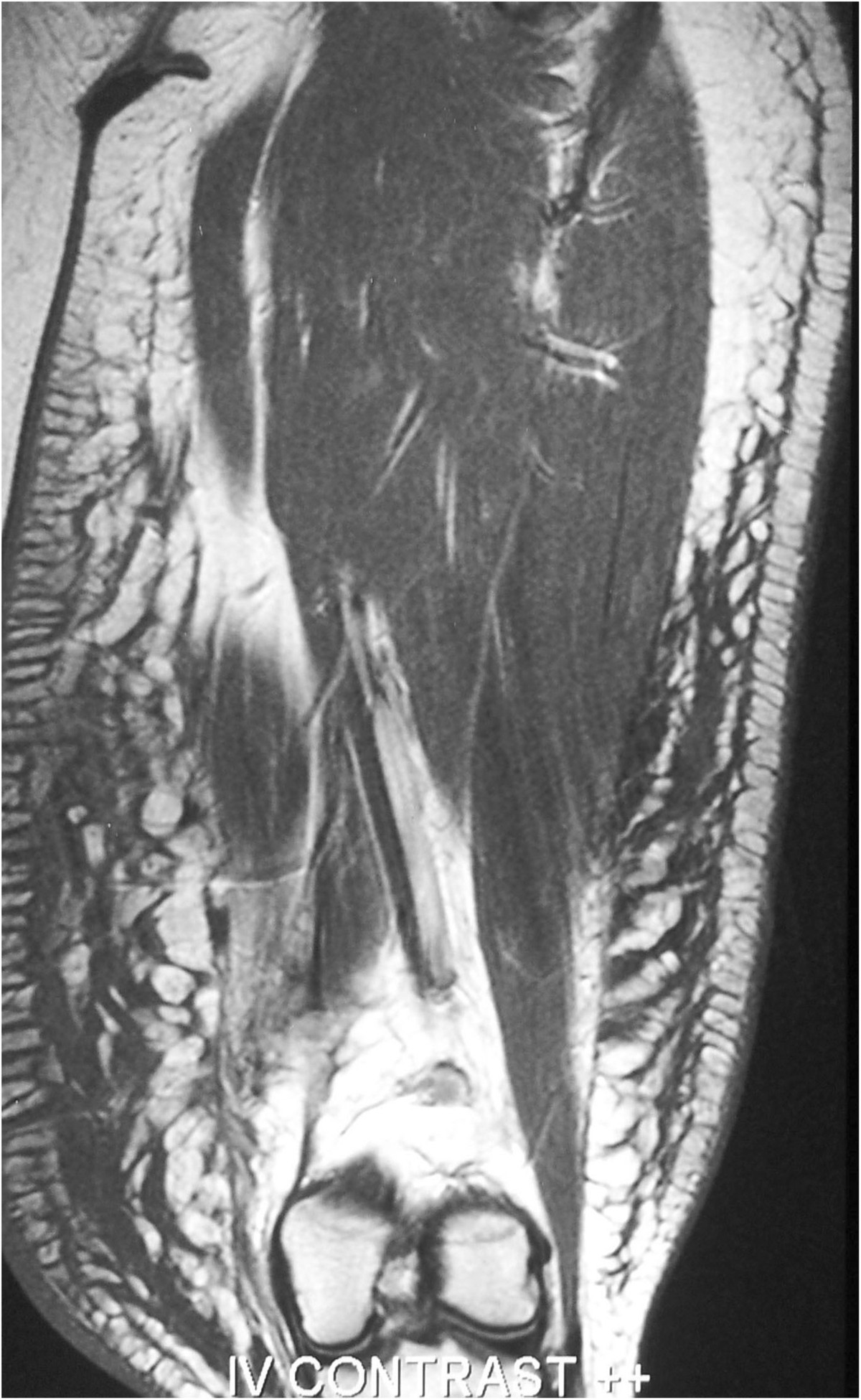
MRI
MRI will display a non-specific, not well-demarcated tumourous mass with variable quantities of fibrous and fatty-tissue .
Signal characteristics
- T1: variable signal intensity with high and low signal areas reflecting fat and fibrous tissue
- T2: variable heterogeneous signal intensity reflecting fat and fibrous tissue
- T1 C+ (Gd): variable enhancement
Radiology report
The radiological report should include a description of the following:
- form, location and size
- tumor margins
- relation to the muscular fascia
- relationship to local nerves and vessels
Treatment and prognosis
The management includes surgical excision with preservation of adjacent neurovascular structures, however, lipofibromatosis is characterized by a high local recurrence rate of up to 70%. There are no known metastases .
History and etymology
Lipofibromatosis was proposed as a separate entity in 2000 by Fetsch et al. , there were earlier descriptions e.g. in 1991 by Grogan et al. and in textbooks .
Differential diagnosis
Conditions which can mimic the appearance of lipofibromatosis include:
- plantar/palmar fibromatosis
- fibrous hamartoma of infancy
- lipoblastoma
Siehe auch:
- Fibrous hamartoma of infancy
- Juvenile aponeurotic fibroma
- Infantile digital fibromatosis
- fibroblastic and myofibroblastic tumors
- EWSRI-SMAD3-rearranged fibroblastic tumor
und weiter:

