mandible
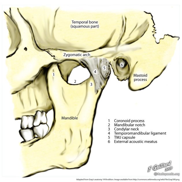
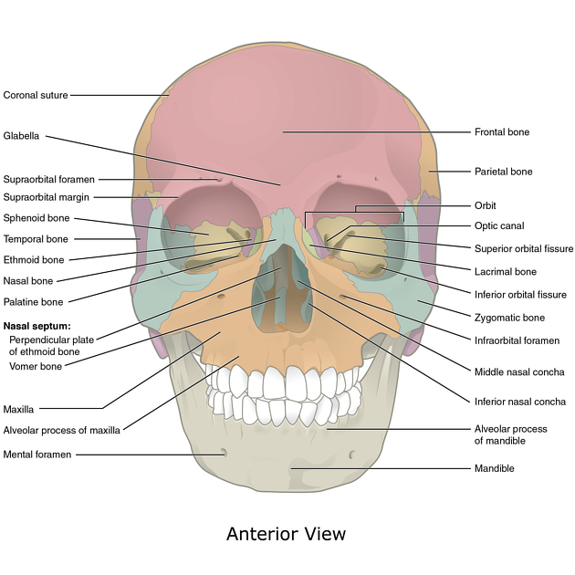
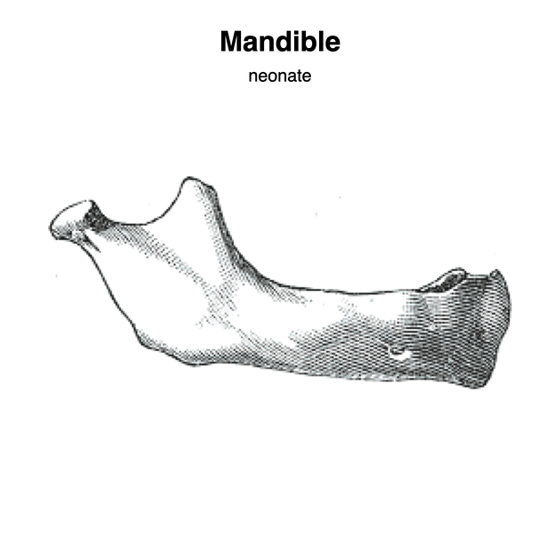
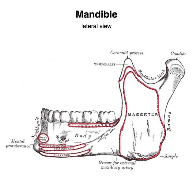
The mandible is the single midline bone of the lower jaw. It consists of a curved, horizontal portion, the body, and two perpendicular portions, the rami, which unite with the ends of the body nearly at right angles (angle of the jaw). It articulates with both temporal bones at the mandibular fossa at the temporomandibular joints (TMJ).
Gross anatomy
Osteology
Body
The body of the mandible is curved, somewhat like a horseshoe, with two surfaces and two borders. The mandibular symphysis is located in the midline, a point of fusion. The parasymphysis extends from the midline to past the canine.
- external surface
- midline ridge indicating the symphysis
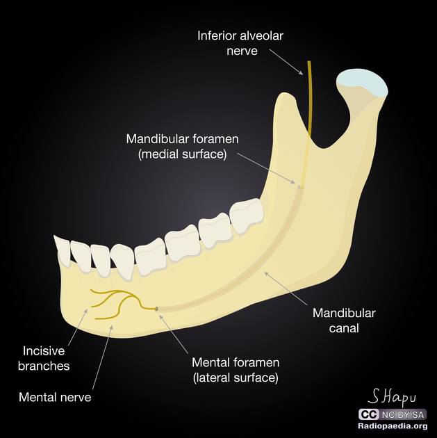
- mental foramen: inferior to second premolar tooth (with normal variants between the canine and 1st molar), midway between the superior and inferior borders; allows for the passage of the mental vessels and mental nerve
- internal surface
- concave from side-to-side
- origin of geniohyoid and genioglossus (from mental spines) and mylohyoid (mylohyoid line)
- fossae for the sublingual and submandibular salivary glands
- superior (or alveolar) border
- wider behind than in front
- hollowed for reception of teeth (normally 16)
- attachment of buccinator muscle
- inferior border
- rounded, longer than the superior border and thicker in front than behind
- groove for the facial artery may be present at the point it joins the ramus of the mandible
Ramus
The ramus is quadrilateral in shape, and has two surfaces, four borders, and two processes and one canal:
- external (or lateral) surface
- flat; gives attachment to the masseter muscle
- inner (or medial) surface
- mandibular foramen: opening of the mandibular canal, which transmit the inferior alveolar vessels and nerve
- lingula of the mandible: prominent, sharp ridge in front of the mandibular foramen; gives attachment to the sphenomandibular ligament
- mandibular foramen: opening of the mandibular canal, which transmit the inferior alveolar vessels and nerve
- lower border
- thick, straight and continuous with the inferior border of the body of the mandible
- posterior border
- thick, smooth, rounded and covered by the parotid gland
- angle of the mandible is at its junction of the posterior border and the body
- medial pterygoid muscle inserts into the medial aspect of the angle of the mandible
- anterior border
- thin above and thicker below; continuous with the oblique line
- upper border
- thin
- consists of the coronoid process anteriorly and the condylar process posteriorly separated by the mandibular notch (a.k.a. sigmoid notch)
The mandibular canal runs obliquely downward and forward in the ramus, and then horizontally forward in the body, where it is placed under the alveoli and communicates with them by small openings. On arriving at the incisor teeth, it turns back to communicate with the mental foramen, giving off two small canals which run to the cavities containing the incisor teeth. It contains the inferior alveolar vessels and nerve, from which branches are distributed to the teeth via the incisive nerve.
Coronoid process
- thin, triangular eminence from the upper border of the ramus of the mandible
- separated from the condylar process posteriorly by the mandibular notch
- temporalis muscle inserts into its medial and lateral surfaces
- masseter muscle also inserts into its lateral surface
Condylar process
Please refer to our article on condylar process of the mandible for a specific discussion
Blood supply
- facial artery (branch of external carotid artery)
- lingual artery (branch of external carotid artery)
- inferior alveolar artery (branch of maxillary artery)
Siehe auch:
- mental foramen
- Unterkieferfraktur
- mandibular foramen
- Schädel
- Kiefergelenk
- Canalis mandibulae
- Processus muscularis
und weiter:
- Arteria carotis externa
- Nervus facialis
- skull
- Keratozystischer odontogener Tumor
- lateral pterygoid muscle
- temporalis muscle
- masticator space
- Franceschetti-Zwahlen-Syndrom
- pterygoid fovea
- Musculus digastricus
- calcifying epithelial odontogenic tumour
- pindborg tumour
- keratocystic odentogenic tumour
- Alveolarfortsatz
- lingula - mandible


 Assoziationen und Differentialdiagnosen zu mandible: