melanotic meningioma
Melanotic meningioma is a rare histological variant of WHO grade I (benign) meningioma.
Epidemiology
As only several dozen cases have been reported in the literature, no significant difference in prevalence has been found between genders/age groups.
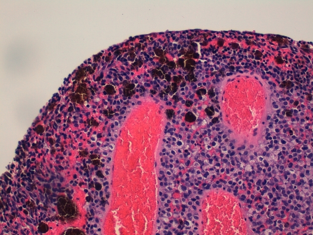
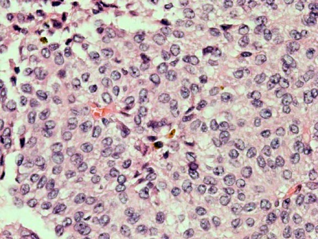
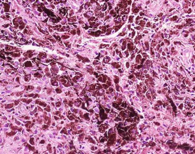
Pathology
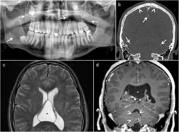
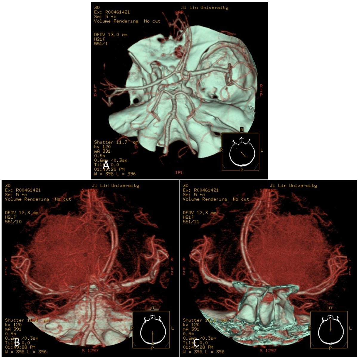
Melanotic meningiomas arise from leptomeningeal melanocytes, small pigment cells found mostly in the meninges covering the ventrolateral surfaces of the medulla oblongata, posterior cranial fossa or Meckel cave .
Radiographic features
Given the small number of reported cases it is difficult to determine the most characteristic appearance.
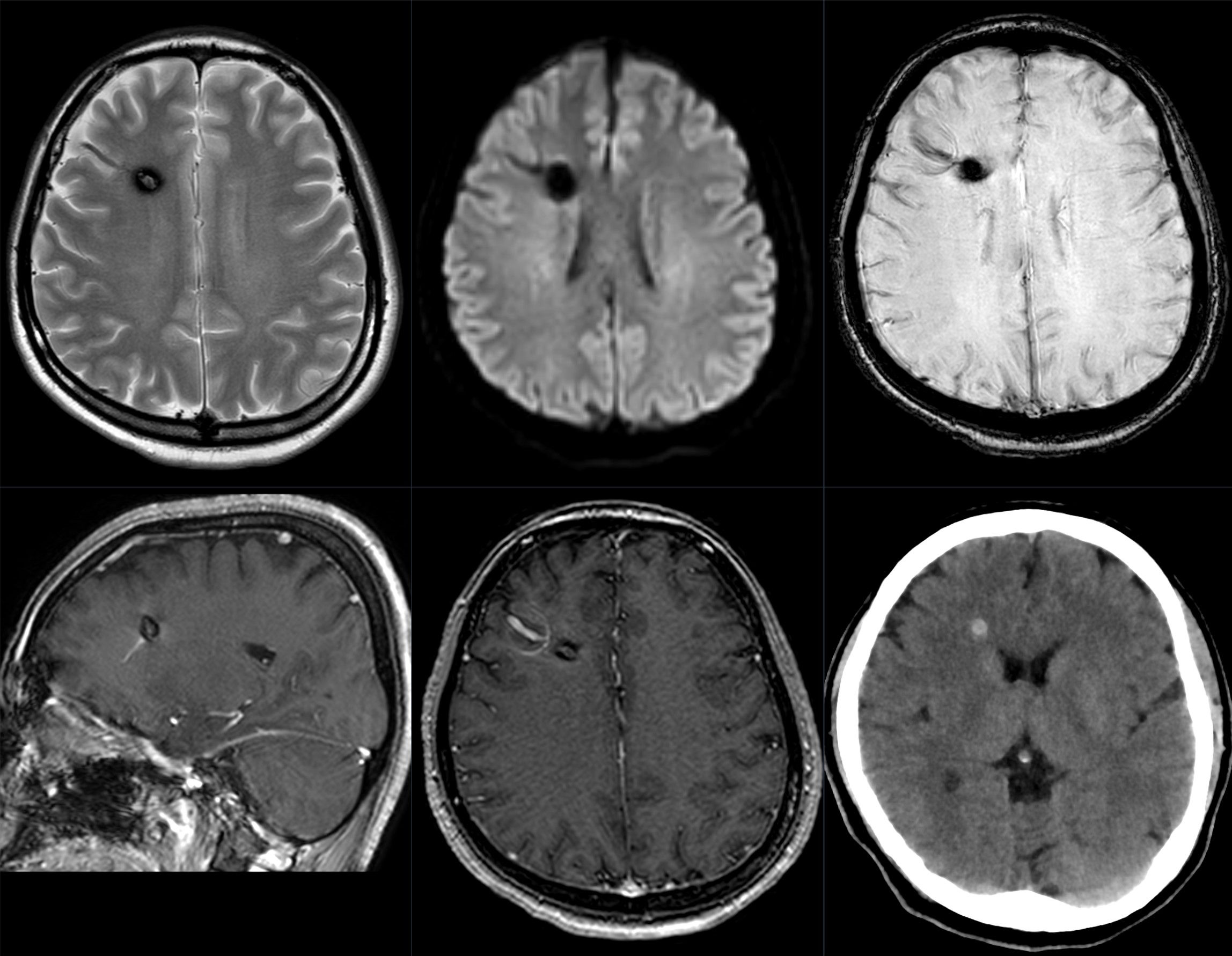
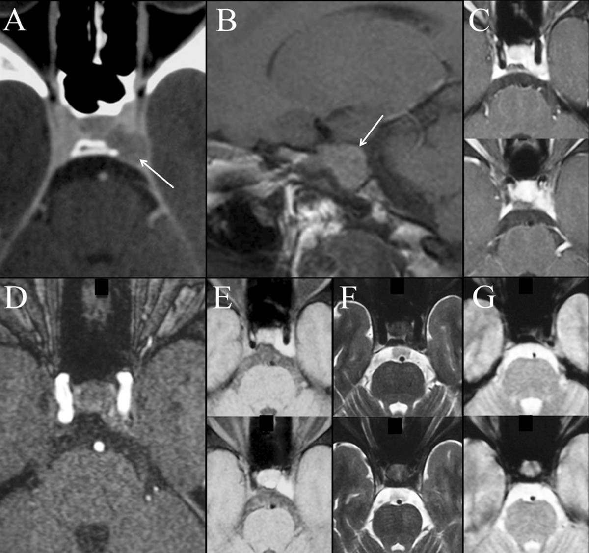
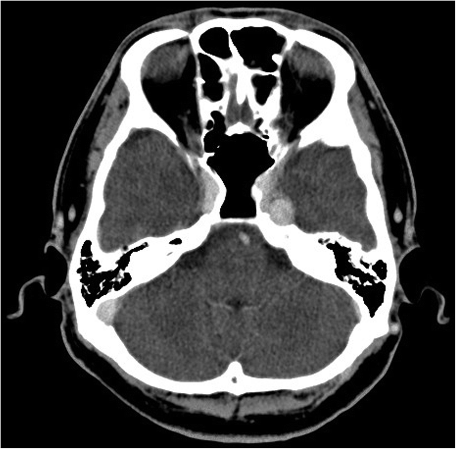
CT
These tumors are most commonly isodense to grey matter, with variable enhancement .
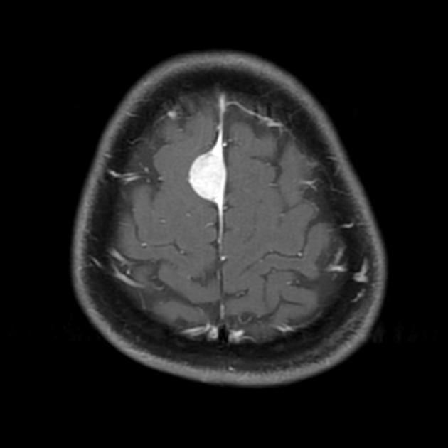
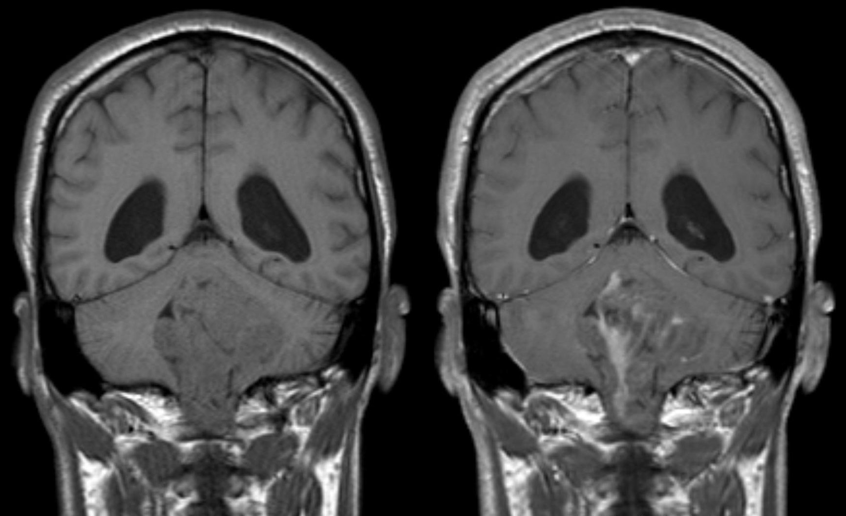
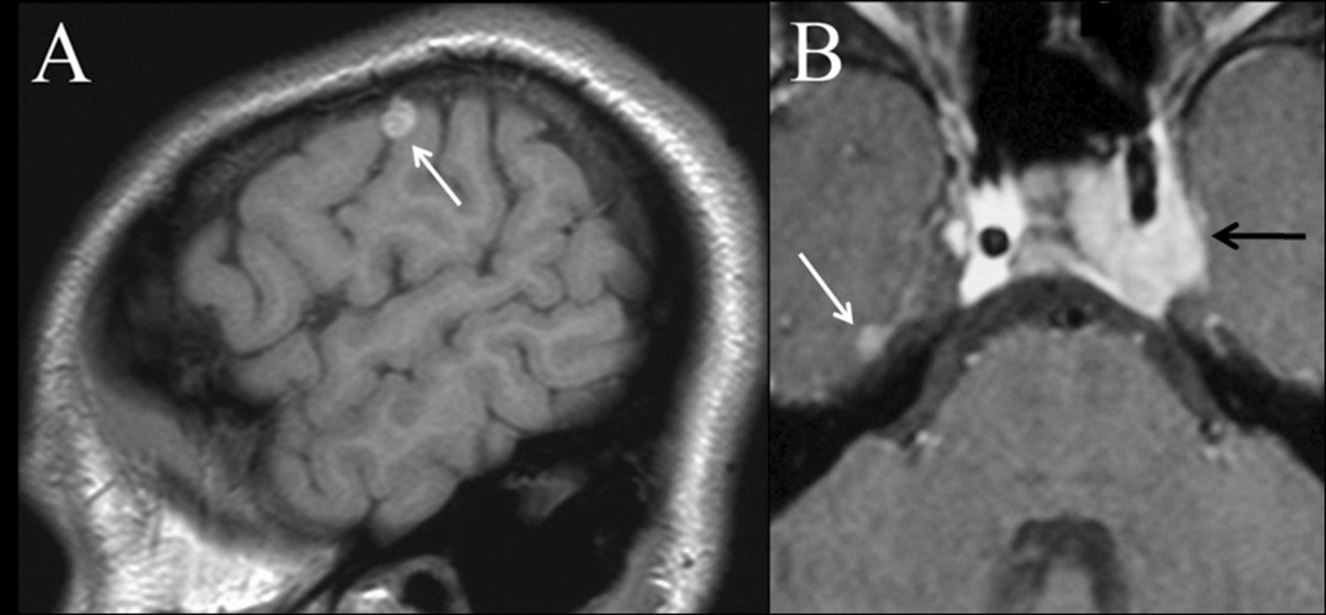
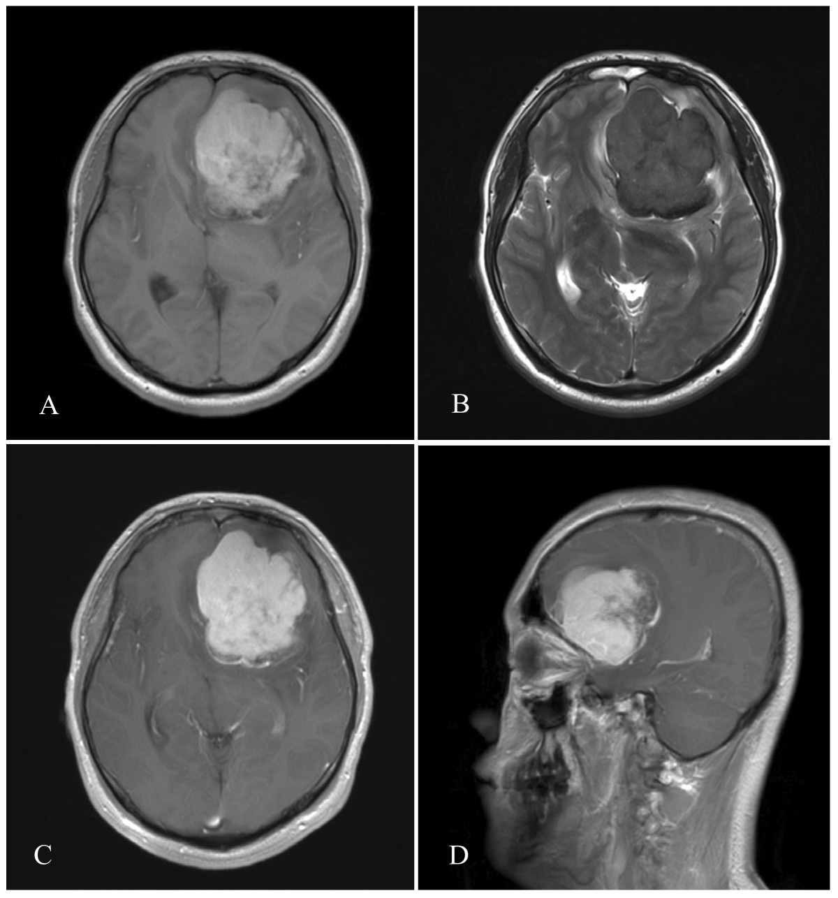
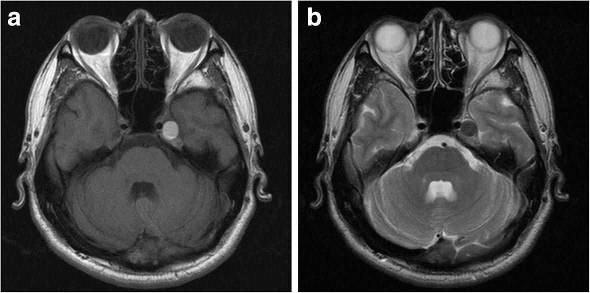
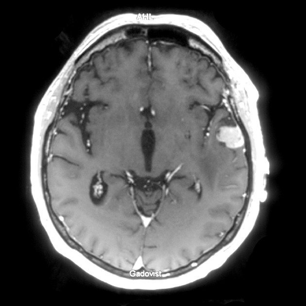
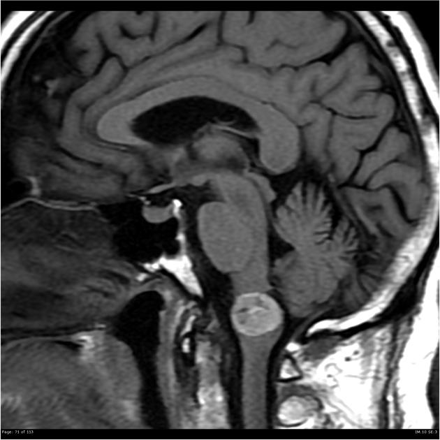
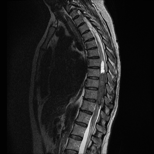
MRI
Different signal intensities are probably correlated with the amount of melanin in the pigment cells. Possible causes of low signal intensity on T2-weighted sequences include decreased water content, paramagnetism, susceptibility effects or hemorrhage .
- T1: iso- or hyperintense
- T2: hypo- or hyperintense
- T1 C+: homogenous enhancement
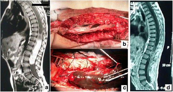
Treatment and prognosis
Similar to other meningiomas the treatment of melanotic meningiomas is usually surgical excision, with complete tumor removal being the best therapeutic option (in terms of disease-free survival) .
Siehe auch:
- Meningeom
- Ependymom
- Astrozytom
- Hämangioblastom
- Kavernom
- metaplastic meningioma
- meningeale Tumoren
- hyperdense intrazerebrale Läsionen
- verkalkte Meningen
und weiter:


 Assoziationen und Differentialdiagnosen zu meningeales Melanozytom: