ösophageales Lipom
Successful
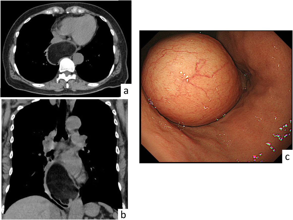
removal of a giant esophageal lipoma by thoracoscopic enucleation: a case report. Preoperative imaging. (a horizontal, b coronal) CT scan of the chest revealed a 10 × 7 cm homogenous mass in the middle and lower esophagus. c Upper endoscopy revealed a submucosal tumor with normal mucosa arising from the left esophageal wall

Submuköses
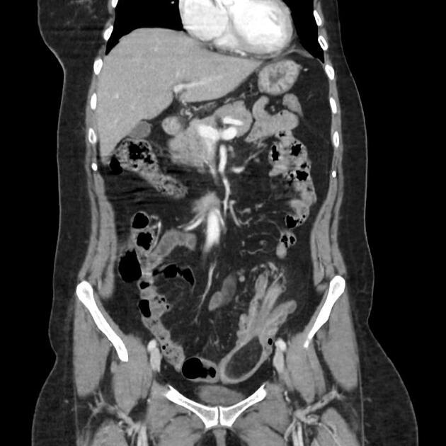
Lipom im unteren Ösophagus bei einem Mann. Computertomographie axial und coronar.

Lipom des
Ösophagus: Glatt begrenzte, submuköse, kleine Formation mit fett-typischen Dichtewerten (-84HU) als Zufallsbefund in der Computertomographie.


 Assoziationen und Differentialdiagnosen zu Lipome des Ösophagus: