prostatic tuberculosis
Prostatic tuberculosis or tuberculous prostatitis is an uncommon extrapulmonary manifestation of Mycobacterium tuberculosis infection.
Epidemiology
Primary tuberculosis of the prostate is rare. Genitourinary tuberculosis contributes to 5-10% of extrapulmonary cases of tuberculosis in developed countries and 15-20% of cases in developing countries. 6.6% of all genitourinary tuberculosis involve the prostate .
Clinical presentation
Patients with tuberculous prostatitis usually present with urethral discharge, painful (sometimes blood-stained) ejaculation, mild ache in the perineum, infertility, and dysuria .
On rectal exam, the prostate may be enlarged in size, firm, and tender. The findings are difficult to differentiate from prostatic hyperplasia and cancer .
Pathology
The possible modes of infective spread include haematogenous, lymphatic, or direct routes.
Radiographic features
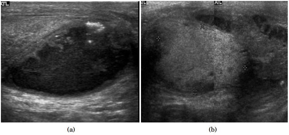
Ultrasound
Findings include :
- enlargement of the prostate gland
- solitary nodular (rare) or multiple hypoechoic areas of variable sizes within the gland
- irregularity of the external contour of the prostate
Transrectal ultrasound (TRUS)
- TRUS shows diffuse hypoechoic lesions within the peripheral zone of the prostate as the typical location is in the peripheral part of the posterior and lateral lobes of the prostate
- color Doppler interrogation shows increased vascularity in the inflammatory phase of granulomatous prostatitis
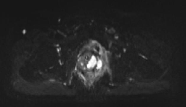
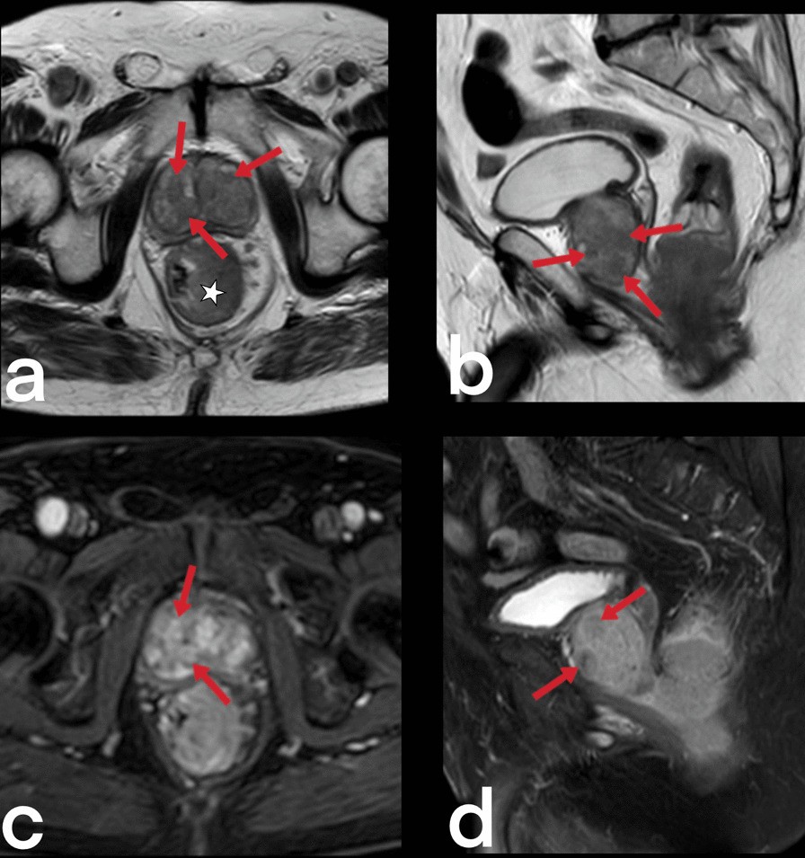
MRI
Characteristic MRI findings of prostatic tuberculosis have not been described due to the relative rarity of cases .
MRI appearance of prostate tuberculosis can be nodular or diffuse :
- nodular type
- T2: characterized by markedly low signal intensity, which is due to the paramagnetic substances such as macrophage-laden oxygen free radicals
- DWI: diffusion restriction
- T1C+: moderate enhancement on dynamic sequences
- diffuse type
- T2: lower signal than that of the normal peripheral zone but not as low as that of nodular lesions. Diffuse streaky areas of low signal intensity on T2 (watermelon skin sign)
Differential diagnosis
Diffuse types may mimic diffuse prostate carcinoma but the citrate level is within normal limits on MRS .
Siehe auch:


 Assoziationen und Differentialdiagnosen zu Tuberkulose der Prostata: