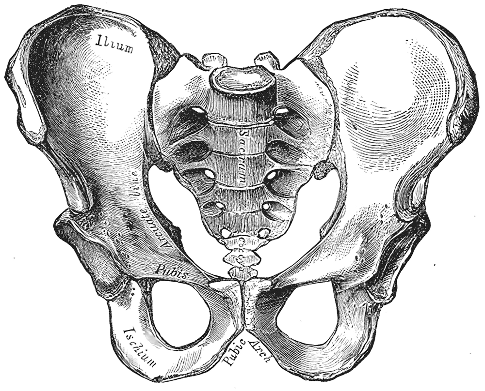
Uterus


The uterus is a hollow, thick-walled, muscular organ of the female reproductive tract that lies in the lesser pelvis.
Gross anatomy
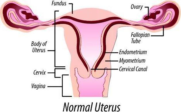
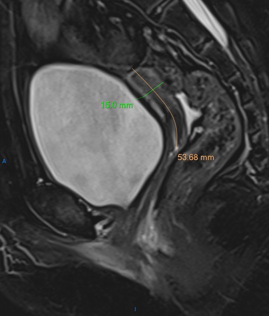
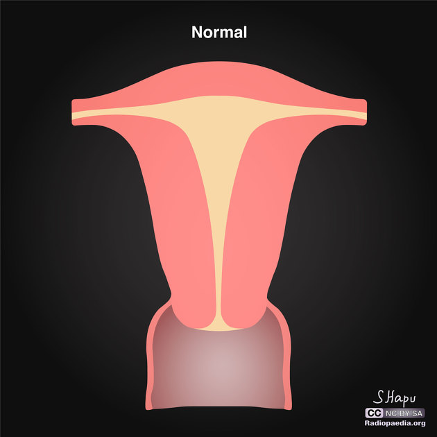
The uterus has an inverted pear shape. In the adult, it measures about 7.5 cm in length, 5 cm wide at its upper part, and nearly 2.5 cm in thickness. It weighs approximately 30-40 grams.
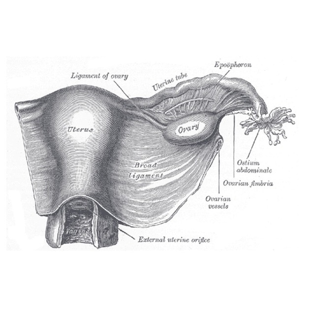
The uterus is divisible into two portions: body and cervix. About midway between the apex and base, is a slight constriction known as the isthmus. The portion above the isthmus is termed the body, and that below, the cervix. The part of the body which lies above a plane passing through the points of entrance of the uterine tubes is known as the fundus.
The body gradually narrows from the fundus to the isthmus. The cavity of the body is a mere slit, flattened anteroposteriorly. It is triangular in shape:
- the base being formed by the internal surface of the fundus between the orifices of the uterine tubes
- the apex by the internal orifice of the uterus through which the cavity of the body communicates with the canal of the cervix
The uterine cervix, although anatomically a part of the uterus, has a different function and is associated with separate pathological entities. It is discussed in detail in a separate article.
Attachments
Musculotendinous and ligamentous
- anterior: pubocervical ligament
- lateral: transverse cervical ligaments (cardinal or Mackenrodt's)
- posterior: uterosacral ligaments
- inferior: puborectalis and pubovaginalis parts of the levator ani muscle
Relations
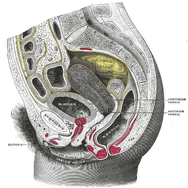
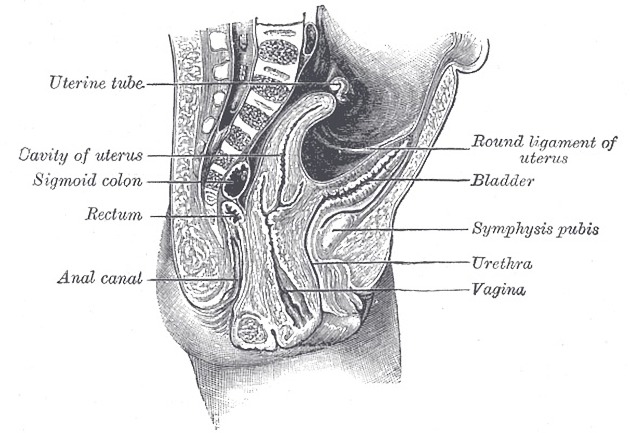
- anteriorly: bladder; uterovesical pouch
- posteriorly: rectum; pouch of Douglas
- laterally: broad ligament; uterine vessels
- uterine tubes open into its upper part
- inferiorly: uterine cavity communicates with that of the vagina
Position
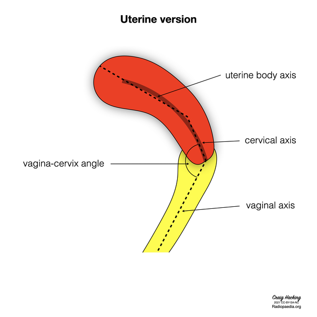
The most common position of the uterus is anteverted (cervix angles forward) and anteflexed (body is flexed forward). The position of the uterus in the adult is liable to considerable variation, depending chiefly on the condition of the bladder and rectum. When the bladder is empty the entire uterus is directed forward and is at the same time bent on itself at the junction of the body and cervix, so that the body lies upon the bladder. As the latter fills, the uterus gradually becomes more and more erect, until with a fully distended bladder the fundus may be directed backward toward the sacrum.
In the fetus, the uterus is contained in the abdominal cavity, projecting beyond the superior aperture of the pelvis. The cervix is considerably larger than the body.
At puberty, the uterus is pyriform in shape and weighs from 14 to 17 g. It has descended into the pelvis, the fundus being just below the level of the superior aperture of this cavity. The palmate folds are distinct and extend to the upper part of the cavity of the organ.
During menstruation, the organ is enlarged, more vascular, and its surfaces rounder; the external orifice is rounded, its labia are swollen, and the lining membrane of the body thickened, softer, and of a darker color.
During pregnancy, the uterus becomes enormously enlarged, and by the eighth month reaches the epigastric region. The increase in size is partly due to growth of pre-existing muscle, and partly to the development of new fibers.
After parturition, the uterus nearly regains its usual size, weighing about 42 g; but its cavity is larger than in the virgin state, its vessels are tortuous, and its muscular layers are more defined. The external orifice is more marked, and its edges present one or more fissures.
In old age, the uterus becomes atrophied, and paler and denser in texture; a more distinct constriction separates the body and cervix. The internal orifice is frequently, and the external orifice occasionally, obliterated, while the lips almost entirely disappear.
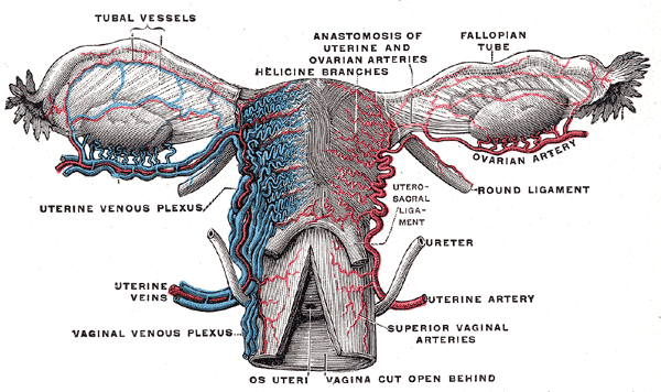
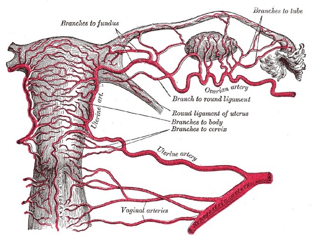
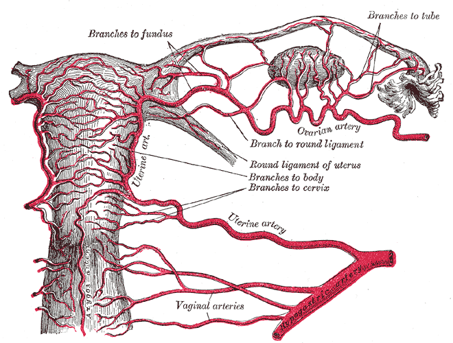
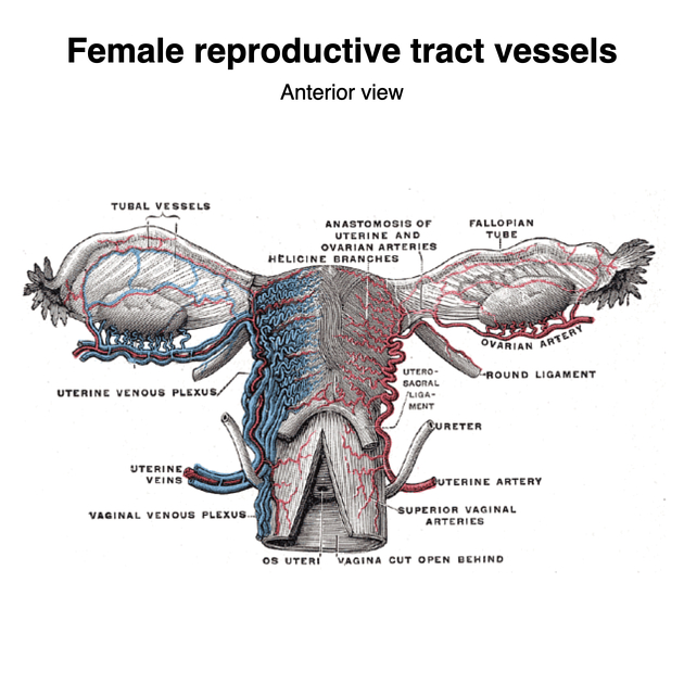
Arterial supply
- uterine arteries and ovarian arteries
- the terminations of the ovarian and uterine arteries unite and form an anastomotic trunk from which branches are given off to supply the uterus
- in the impregnated uterus, the arteries carry the blood to the intervillous space of the placenta
Venous drainage
- uterine vein draining into internal iliac vein
- in the impregnated uterus, the veins convey blood away from the intervillous space of the placenta
Lymphatic drainage
- fundus: para-aortic nodes
- body/cervix: internal and external iliac nodes; superficial inguinal nodes (via round ligament)
Innervation
The nerves are derived from the hypogastric and ovarian plexuses, and from the third and fourth sacral nerves.
Variant anatomy
Radiographic features
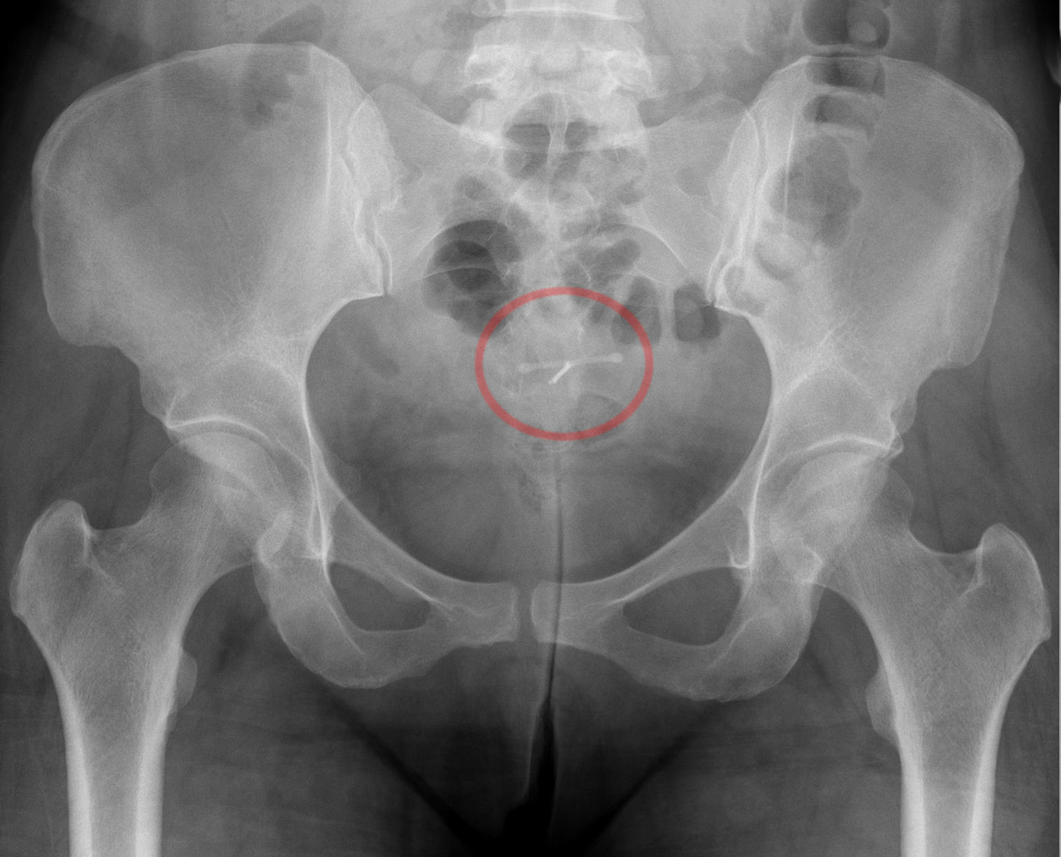
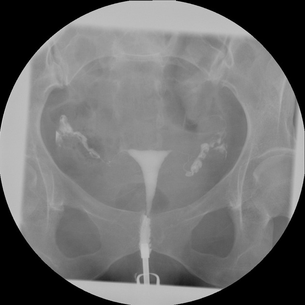
Fluoroscopy
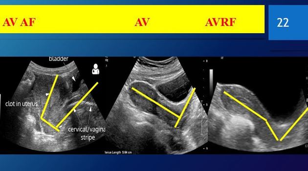
Ultrasound
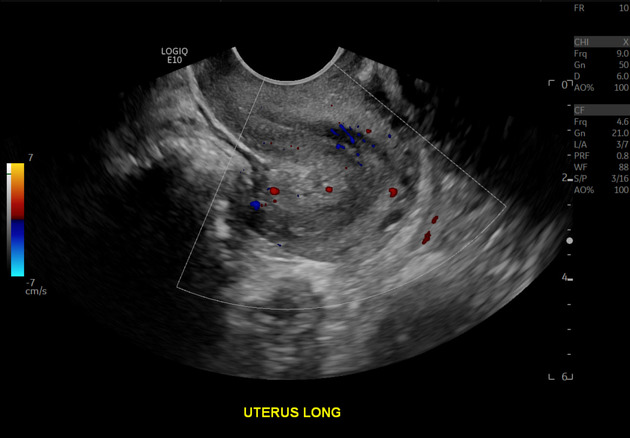
Transabdominal US allows evaluation of the size and position of the uterus in the pelvic cavity. Transvaginal US allows the internal structure of the uterus to be examined .
CT
The uterus appears as a homogeneous soft tissue mass posterior to the bladder. It normally enhances post intravenous contrast .
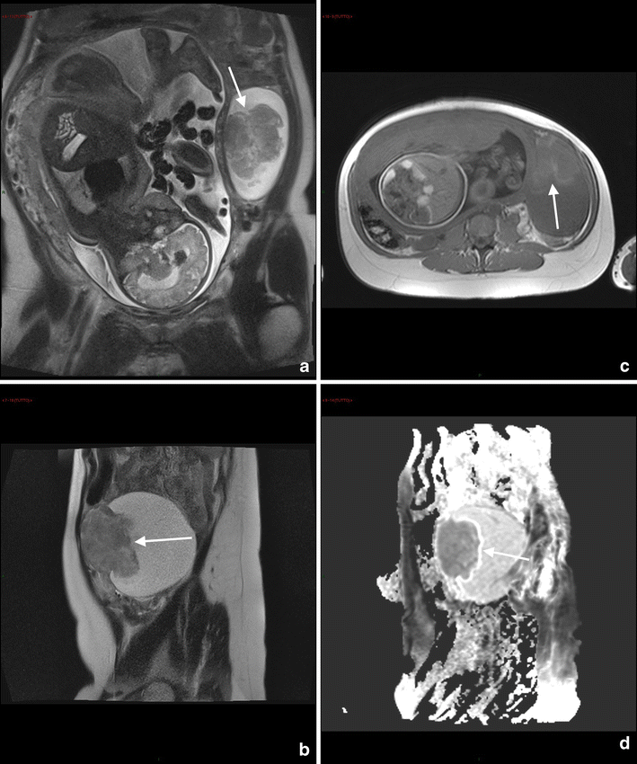
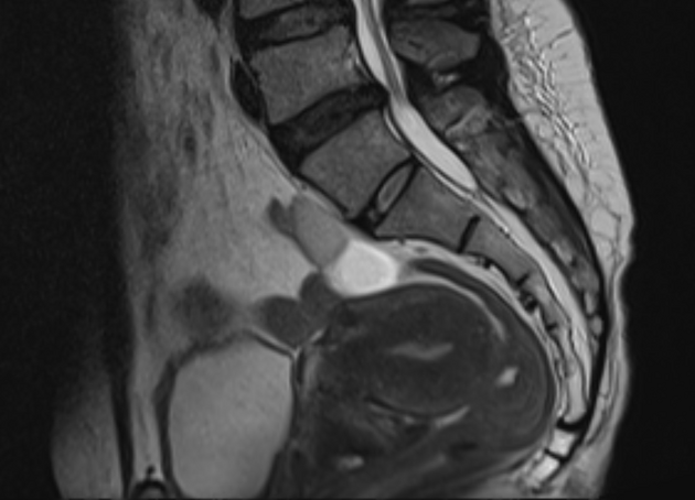
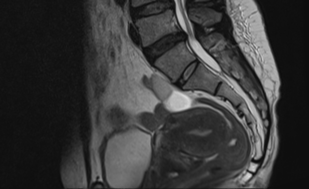
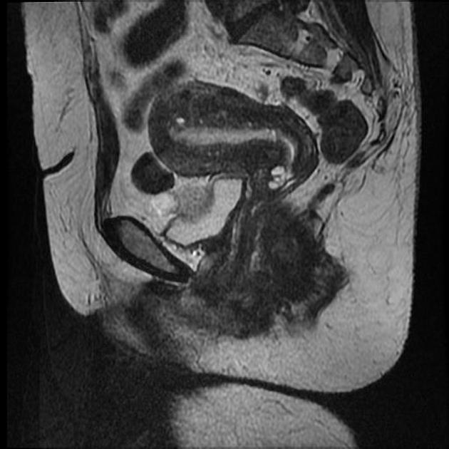
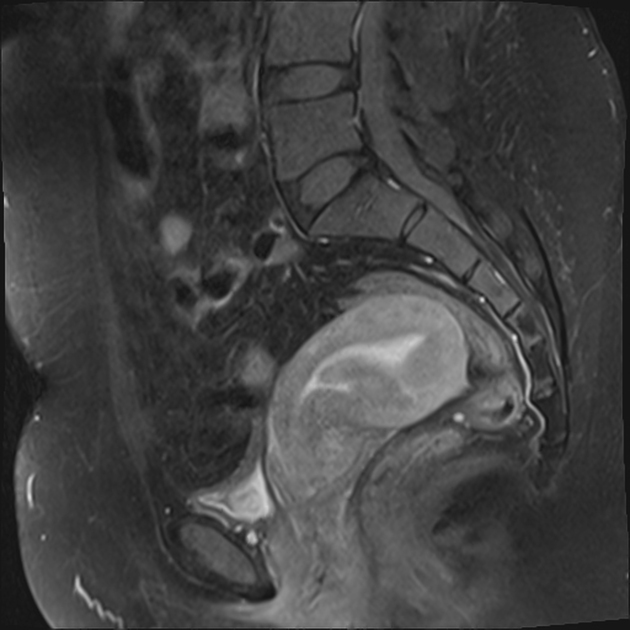
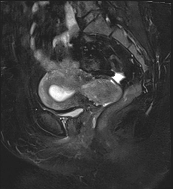
MRI
MRI displays the zonal anatomy of the uterus. The high T2 signal endometrium is outlined by the low T2 signal inner myometrium, known as the junctional zone. The outer myometrium is of intermediate T2 signal. The myometrial layers are indistinguishable on T1 imaging.
There is some physiologic variability to the myometrial zonal appearance. The junctional zone is less distinct pre-menarche and during pregnancy . In the postmenopausal patient, the outer myometrium is thinner and of lower signal due to reduced fluid content and therefore approximates the junctional zone, with poor delineation of the margin in some patients.
Myometrial zonal anatomy has diagnostic implications in the assessment of adenomyosis, and in the staging of endometrial carcinoma, where the depth of myoinvasion is assessed in relation to the junctional zone.
Endometrium on MRI undergoes expected physiological variation in thickness, but the structural, cyclical changes seen on ultrasound are not reproduced.
Related pathology
- neoplastic
- non neoplastic
See also
Siehe auch:
- Leiomyofibrom Uterus
- Becken
- Cervix
- Intrauterinpessar
- Hysterosalpingographie
- ovarian artery
- Fehlbildungen der Gebärmutter
- uterine anomalies
- uterine arterial Doppler ultrasound assessment
- Uteruskarzinom
und weiter:
- Salpinx
- Vagina
- obstetric curriculum
- Colon sigmoideum
- nuchal translucency
- uterine artery
- ovaries
- Müllerian duct anomaly classification
- Harnblase
- knöchernes Becken
- Müller-Gang
- pelvic peritoneal space
- Uterusmyom-Embolisation
- gynäkologisch radiologisches Curriculum
- Ovar
- Uterus in menstruationem
- Embolisation Arteria uterina


 Assoziationen und Differentialdiagnosen zu Uterus: