extramedullary hematopoiesis

Extramedullary
hematopoiesis of the falx. High signal lesion centered on the falx entering the sulci

Extramedullary
hematopoiesis of the falx. Diffuse and intense high signal of the lesion
Extramedullary
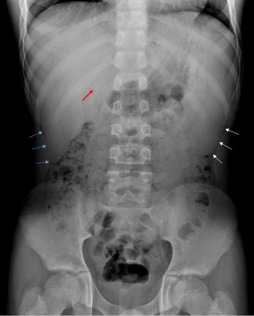
hematopoiesis • Extramedullary hematopoiesis - beta-thalassemia major - Ganzer Fall bei Radiopaedia
Extramedullary
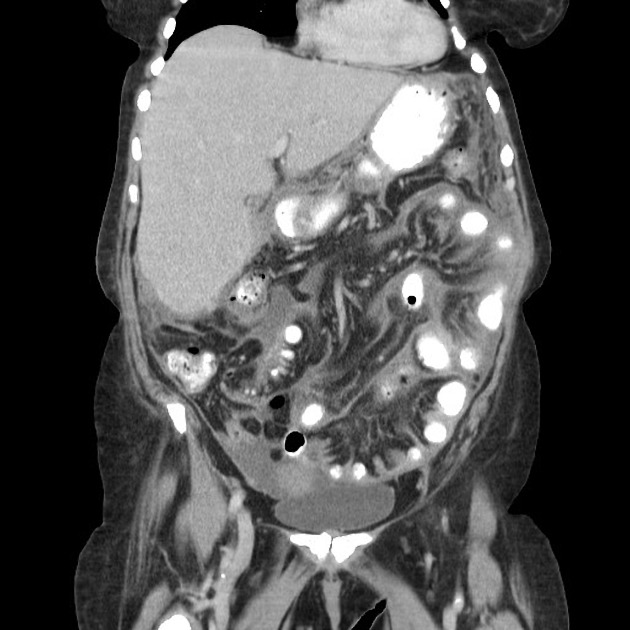
hematopoiesis • Peritoneal extramedullary hematopoesis - Ganzer Fall bei Radiopaedia
Extramedullary
hematopoiesis • Thoracic extramedullary hematopoiesis - Ganzer Fall bei Radiopaedia
Extramedullary
hematopoiesis • Extramedullary hematopoiesis - Ganzer Fall bei Radiopaedia
Extramedullary
hematopoiesis • Thalassemia - Ganzer Fall bei Radiopaedia
Extramedullary
hematopoiesis • Paraspinal extramedullary haematopoesis - Ganzer Fall bei Radiopaedia
Extramedullary
hematopoiesis • Extramedullary hematopoiesis - spleen - Ganzer Fall bei Radiopaedia
Extramedullary
hematopoiesis • Extramedullary hematopoiesis - presacral mass - Ganzer Fall bei Radiopaedia
Extramedullary
hematopoiesis • Extramedullary hematopoiesis - presacral mass - Ganzer Fall bei Radiopaedia
Extramedullary
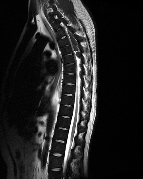
hematopoiesis • Extramedullary hematopoiesis - spinal epidural lesions - Ganzer Fall bei Radiopaedia
Extramedullary
hematopoiesis • Extramedullary hematopoiesis - presacral soft tissue mass - Ganzer Fall bei Radiopaedia
Extramedullary
hematopoiesis • Adrenal extramedullary hematopoiesis - Ganzer Fall bei Radiopaedia
Extramedullary
hematopoiesis • Extramedullary hematopoiesis - pelvic masses - Ganzer Fall bei Radiopaedia
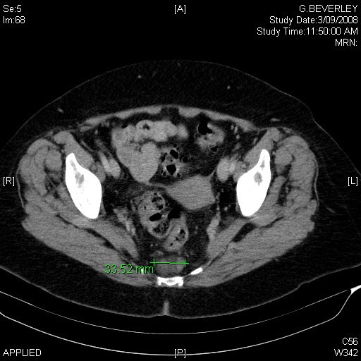
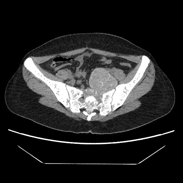
An
interesting diagnosis for a presacral mass: case report. CT transverse view of lesion showing 33.52 mm diameter lesion in the presacral area.
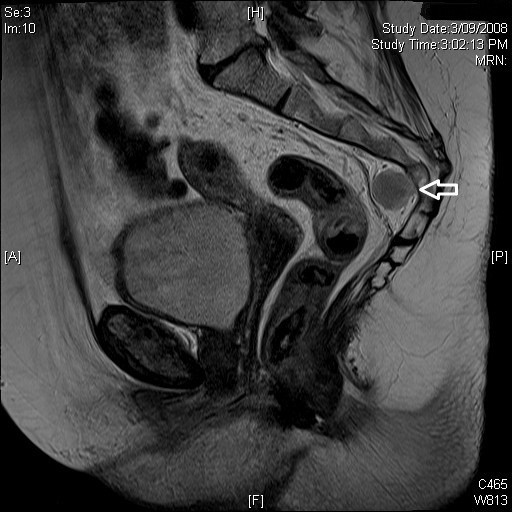
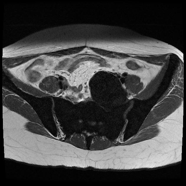
An
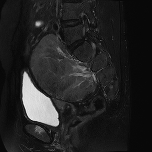
interesting diagnosis for a presacral mass: case report. MRI Sagittal view of lesion.
Extramedullary
hematopoiesis • Extramedullary hematopoiesis - adrenal - Ganzer Fall bei Radiopaedia
Extramedullary
hematopoiesis • Extramedullary haemopoiesis - Ganzer Fall bei Radiopaedia
Extramedullary
hematopoiesis • Extramedullary hematopoiesis - Ganzer Fall bei Radiopaedia

Intrathoracic
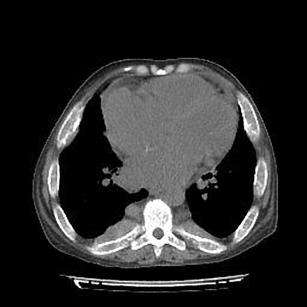
extramedullary haematopoiesis and skeletal involvement in a case of thalassaemia intermedia. Chest axial image shows diffuse structural remodelling of bones with destruction of trabeculae and multiple cortical interruptions; ribs appear widened (arrow).
Extramedullary
hematopoiesis • Thalassemia with extramedullary hematopoiesis - Ganzer Fall bei Radiopaedia
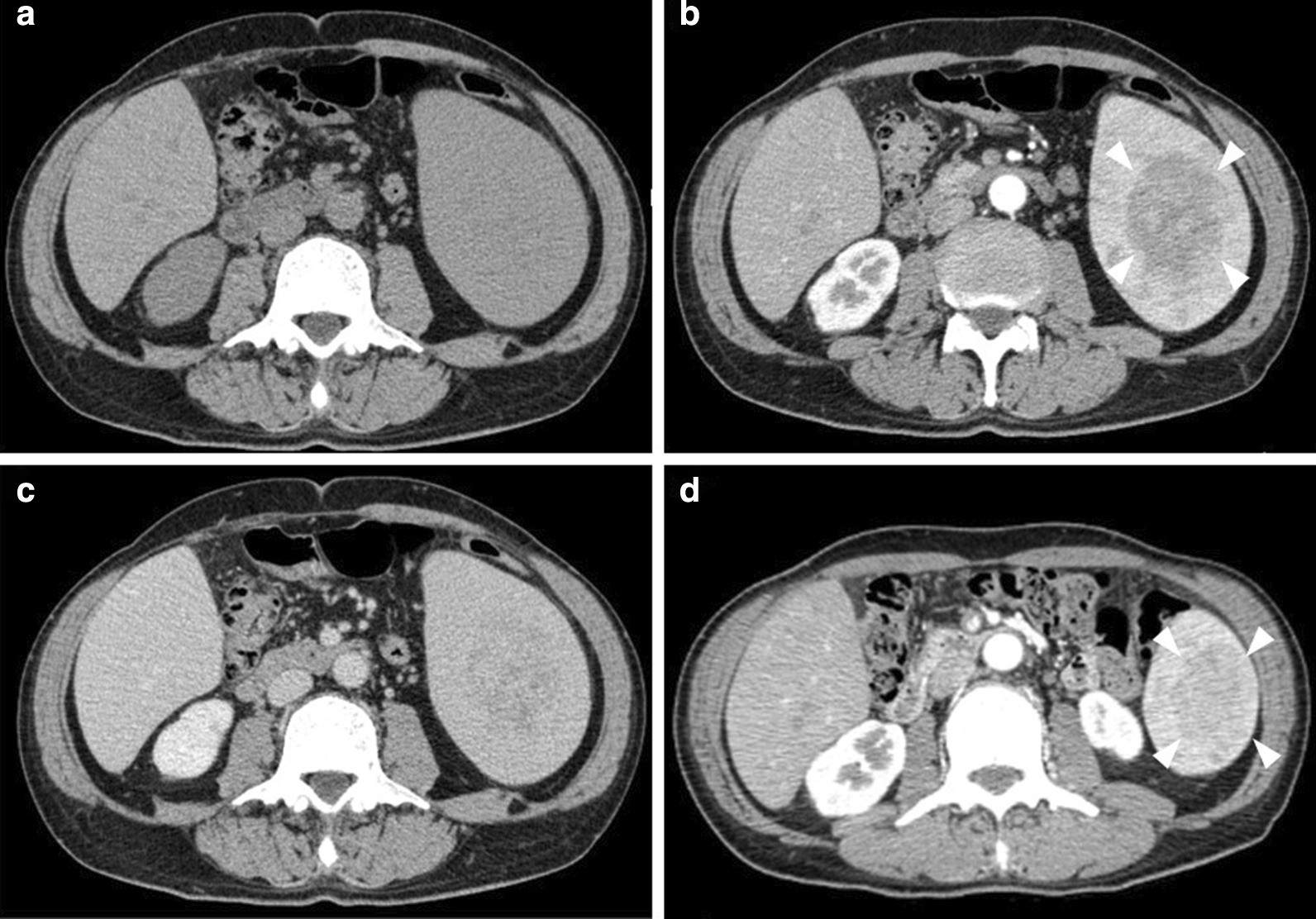
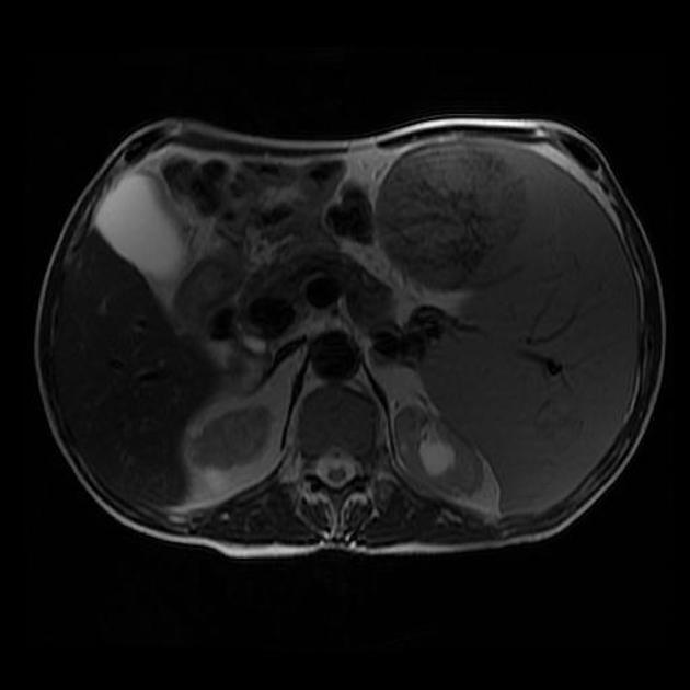
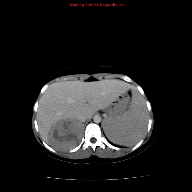
A focal
extramedullary hematopoiesis of the spleen in a patient with essential thrombocythemia presenting with a complicated postoperative course: a case report. Computed tomography scan of the tumor. A computed tomography scan on admission showed a 70 × 45 mm low-density tumor (a). The tumor exhibited heterogeneous hypo-enhancement during the arterial phase (b arrowheads), and thereafter, the enhancement became gradually homogeneous in the equilibrium phase (c). The tumor was 45 × 30 mm in diameter on a preoperative CT scan 6 months before the surgery (d arrowheads)

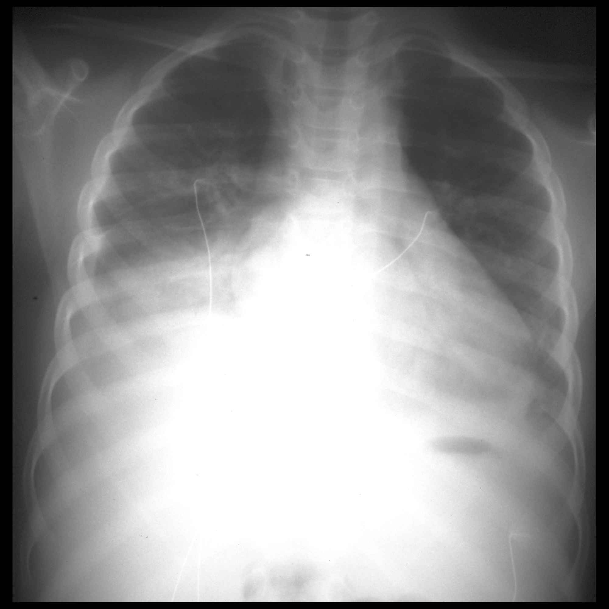
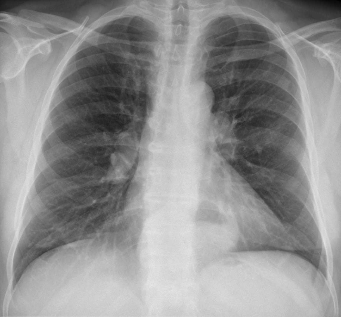
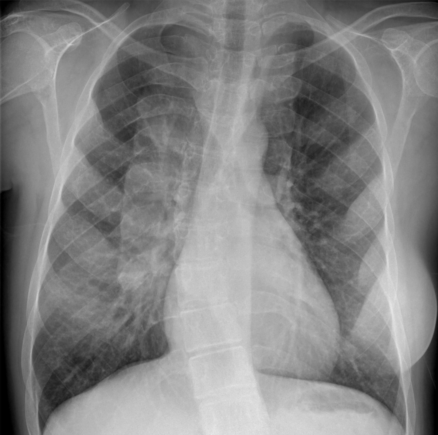
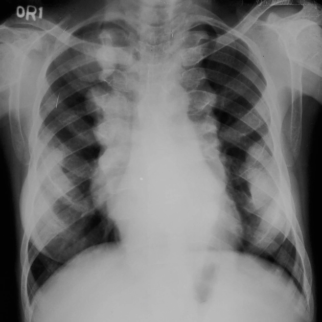
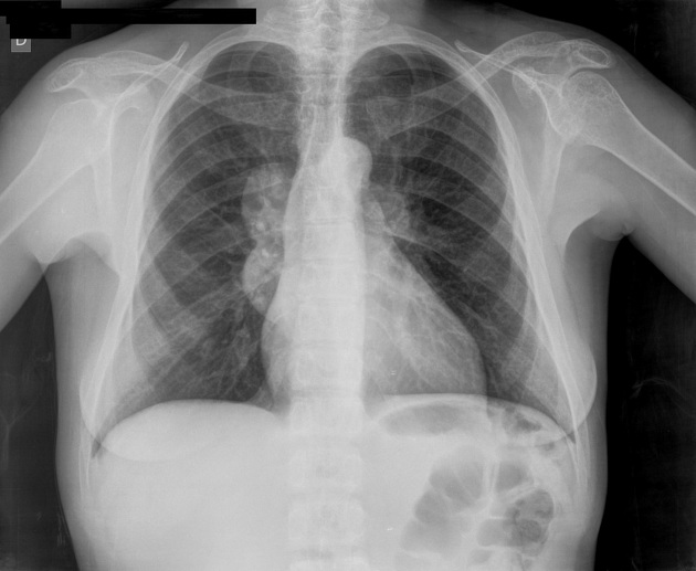
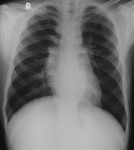
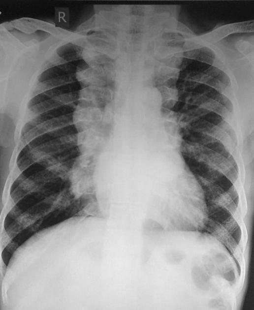
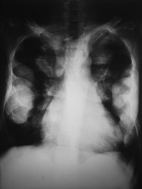
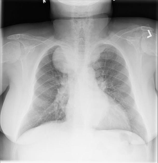
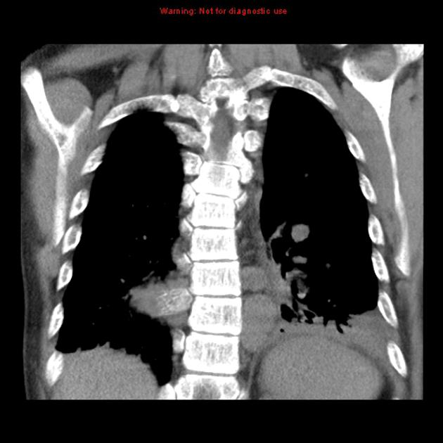
Intrathoracic
extramedullary haematopoiesis and skeletal involvement in a case of thalassaemia intermedia. Chest radiograph shows bilateral well-defined opacities overlying the spine (yellow arrows) and right basal peripheral opacity abutting the costal arches (green arrow).

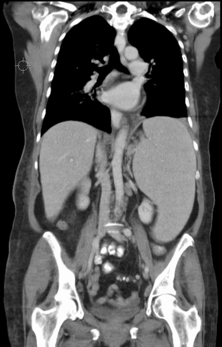
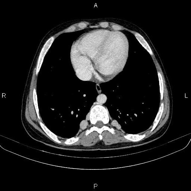
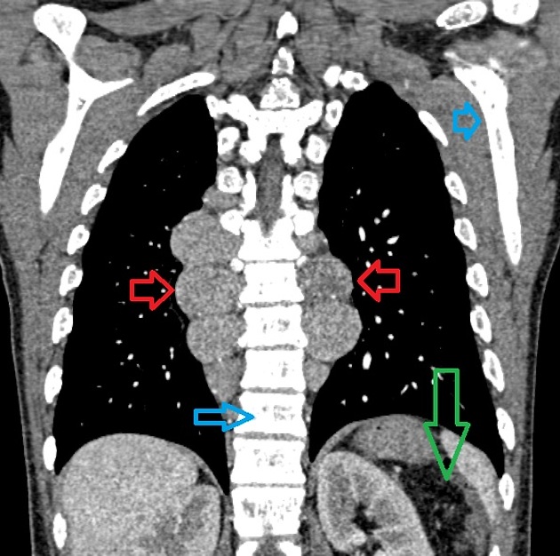
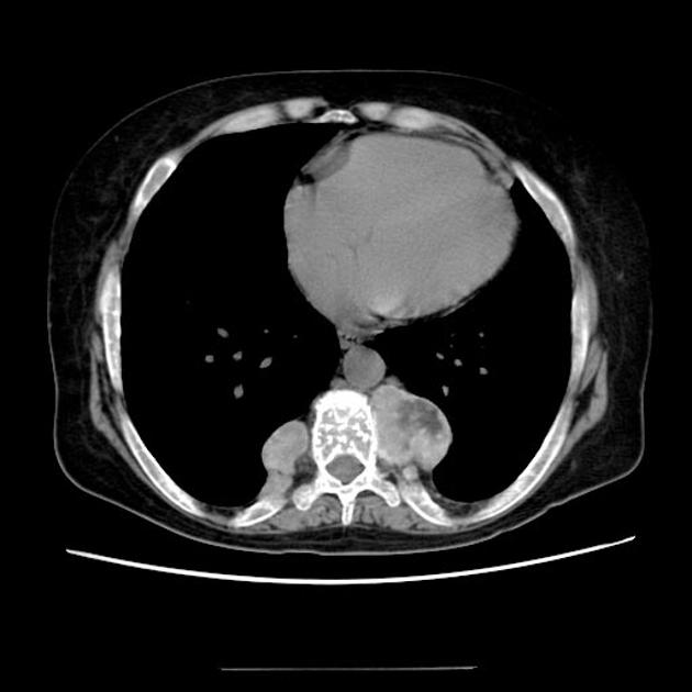
Intrathoracic
extramedullary haematopoiesis and skeletal involvement in a case of thalassaemia intermedia. Chest axial image shows bilateral paravertebral well-marginated soft tissue masses, demonstrating mild enhancement after intravenous injection of contrast medium; diffuse decreased density of bones with structural remodelling is also seen.

Intrathoracic
extramedullary haematopoiesis and skeletal involvement in a case of thalassaemia intermedia. Chest axial image shows bilateral paravertebral well-marginated soft tissue masses and soft tissue in parasternal location (arrow); diffuse decreased density of bones with structural remodelling is also seen.

Intrathoracic
extramedullary haematopoiesis and skeletal involvement in a case of thalassaemia intermedia. Chest axial image shows bilateral paravertebral and right pericostal well-marginated masses, demonstrating mild enhancement after intravenous injection of contrast medium; diffuse decreased density of bones with structural remodelling is also seen.

Intrathoracic
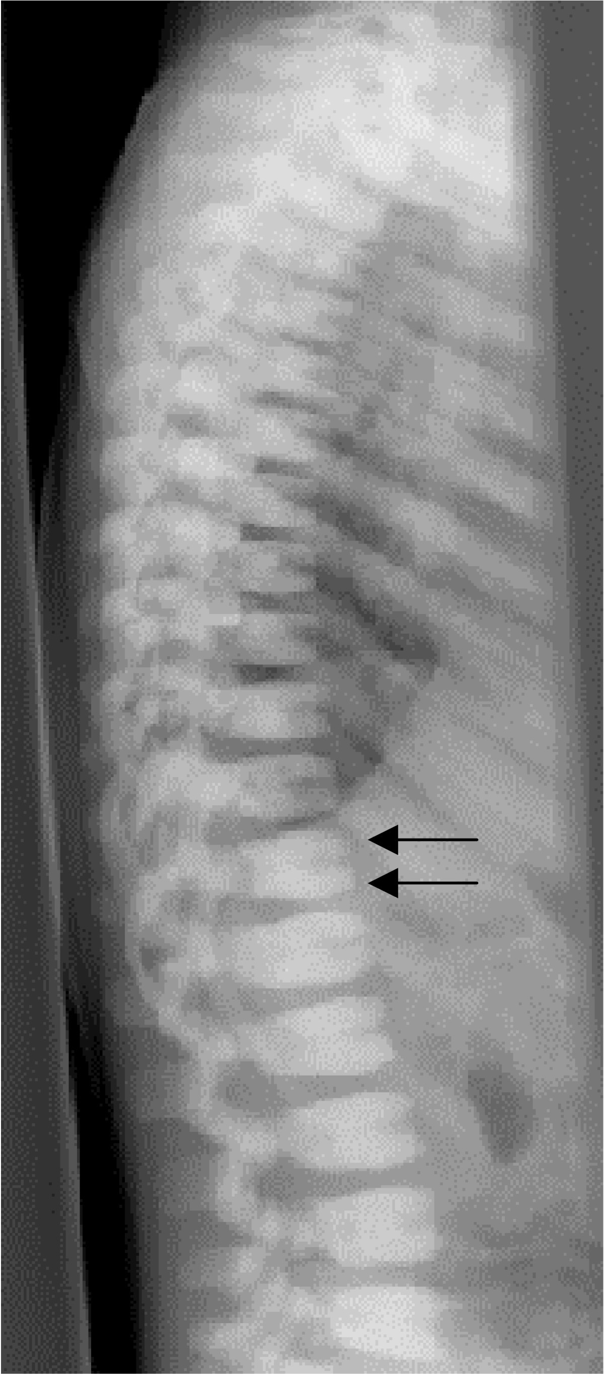
extramedullary haematopoiesis and skeletal involvement in a case of thalassaemia intermedia. Chest axial image shows diffuse structural remodelling of vertebral bodies with trabecular thinning.
Extramedullary
hematopoiesis • Extramedullary hematopoiesis - Ganzer Fall bei Radiopaedia

Intrathoracic
extramedullary haematopoiesis and skeletal involvement in a case of thalassaemia intermedia. Chest CT sagittal image shows multiple cortical interruptions of the sternum (arrows), which appears surrounded by soft tissue.

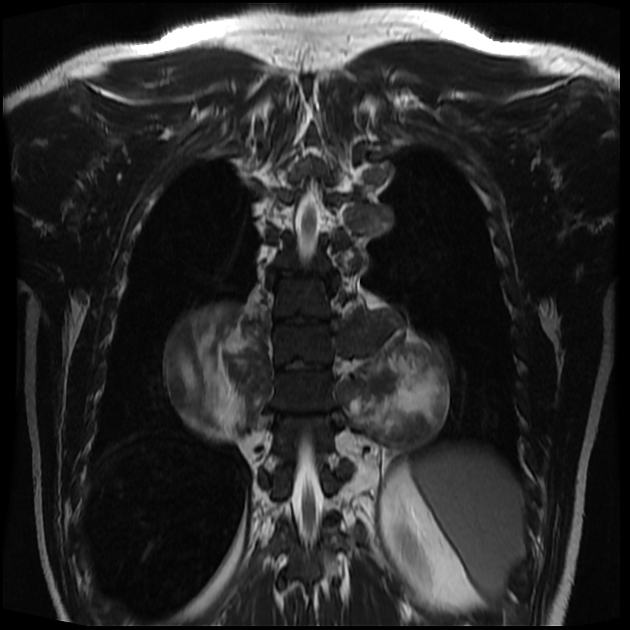
Tumor and
tumorlike conditions of the pleura and juxtapleural region: review of imaging findings. Diagnosis: extramedullary hematopoiesis. Technique: chest CT and MRI. Description: A 42-year-old woman with sickle cell anemia was referred for a routine control examination. The axial CT in the mediastinal window setting (a) shows as incidental findings, two well-delineated paravertebral masses with a homogeneous hypodense aspect. There is no locoregional bone invasion. On both the axial T1 vibe MRI sequence with FS (b) and the axial T2 haste MRI sequence (c), the lesions have the same signal intensity as bone marrow. There is no contrast enhancement of the lesions on the axial T1 vibe sequence with FS (d)
Extramedullary
hematopoiesis • Extramedullary hematopoiesis - Ganzer Fall bei Radiopaedia
Extramedullary
hematopoiesis • Extramedullary hematopoiesis - Ganzer Fall bei Radiopaedia
Extramedullary
hematopoiesis • Extramedullary hematopoiesis - thalassemia - Ganzer Fall bei Radiopaedia

Extramedullary
hematopoiesis of the falx. Slightly high signal mass along the falx cerebri
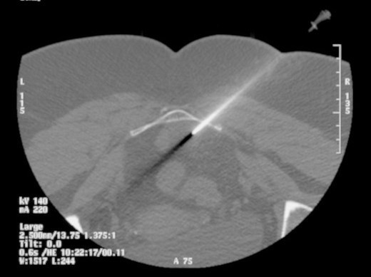
An
interesting diagnosis for a presacral mass: case report. CT guided biopsy through presacral lesion using posterolateral approach.
Extramedullary hematopoiesis is a response to the failure of erythropoiesis in the bone marrow.
This article aims to a general approach on the condition, for a dedicated discussion for a particularly involved organ, please refer to the specific articles on:
Extramedullary hematopoiesis usually affects visceral organs like liver, spleen, lymph nodes and involves thorax. Less commonly it can affect the pleura, lungs, gastrointestinal tract, breast, skin, brain, kidneys, and adrenal glands.
Pathology
Etiology
Radiological features
- most common: diffuse visceromegaly (splenomegaly and hepatomegaly)
- best evaluated with ultrasound, CT or MRI
- lesions are typically hypermetabolic, hence FDG-18 PET avid
- rarely, can result in focal masses in liver and spleen that need to be differentiated from malignancy
- most common intrathoracic finding is a posterior mediastinal mass
- may be either unilateral or bilateral
- smooth, sharply-delineated, often lobulated margins
- fat can be seen, if chronic
- calcification is very atypical
- other than this, within the thorax, there can be rib expansion and rarely pulmonary infiltrates
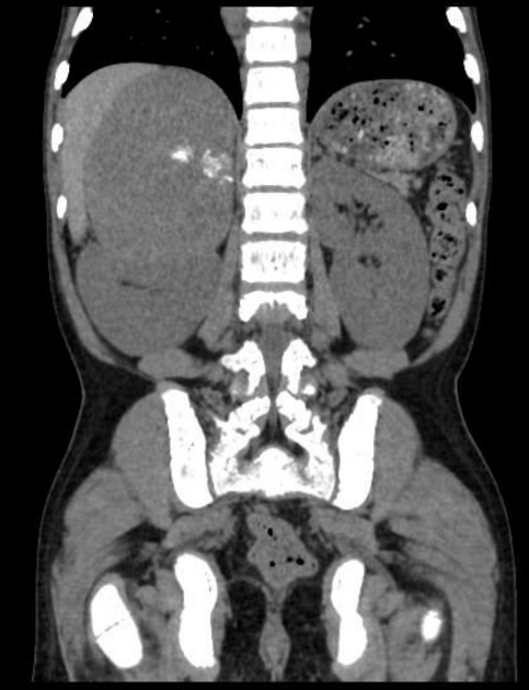
- perirenal soft tissue with normal renal contour can be seen (mimicking lymphoma or Erdheim-Chester disease like appearance) . It has been found to be the most common retroperitoneal finding
- focal or diffuse peritoneal nodules can be seen
- can present as pre-sacral soft tissue mass
- epidural soft tissue masses with peripheral fat can be seen in spinal cord or CNS with compression of spinal cord
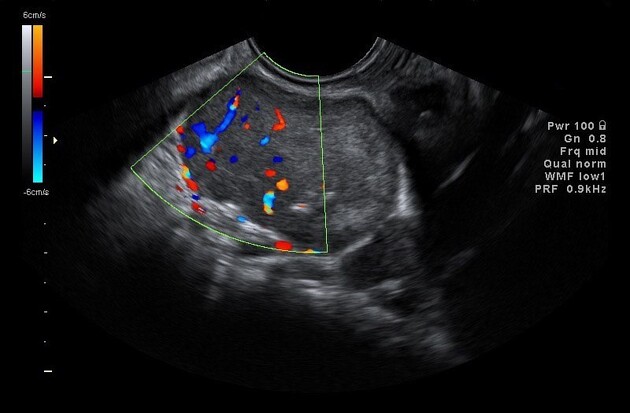
- These masses are generally hypervascular with high chances of occurence of bleeding as a complication of biopsy. Hence, avoid biospy near vital structures like spinal cord to avoid risk of spinal cord compression. FNAC is a better option at such sites .
- For treatment, giving radiotherapy on the involved site or excision of the mass or multiple blood transfusions to decrease extramedullary hematopoiesis can be done .
Siehe auch:
- Splenomegalie
- Osteomyelofibrose
- Sichelzellenanämie
- Tumoren des hinteren Mediastinums
- Thalassämie
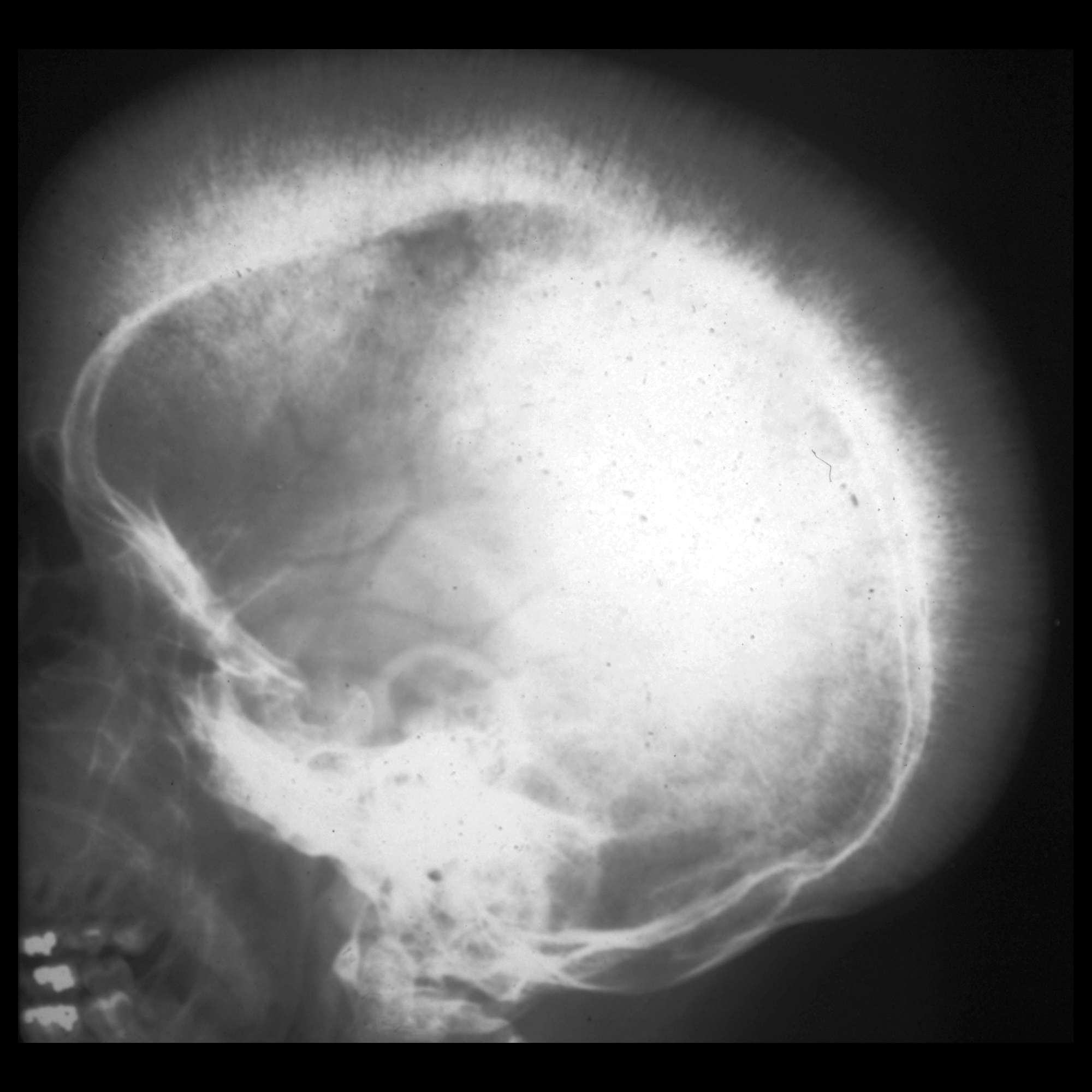
- Bürstenschädel
- Knochenmarksexpansion
- paraspinal mass
- extraossäres Plasmozytom
- thoracic paraspinal mass
- extramedulläre Blutbildung intrakraniell
- Hereditäre Sphärozytose
- diffuse skelettale Sklerosierung
und weiter:
- epidurale intraspinale Raumforderung
- posterior mediastinal masses
- paravertebrale Raumforderungen
- spinale Epiduralblutung
- Abhebung Ligamentum longitudinale posterius
- adrenal extramedullary haematopoietic tumour
- intraspinal extramedullary haematopoiesis
- peritoneal extramedullary hematopoesis
- extramedulläre Hämatopoese in der Milz
- muskuloskelettale Manifestationen bei Sichelzellanämie
- präsakrales Myelolipom
- mediastinale extramedulläre Hämatopoese
- präsakrale extramedulläre Hämatopoese


 Assoziationen und Differentialdiagnosen zu extramedulläre Hämatopoese: