breast MRI enhancement curves
Breast MRI
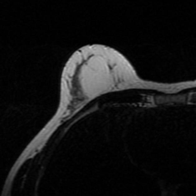
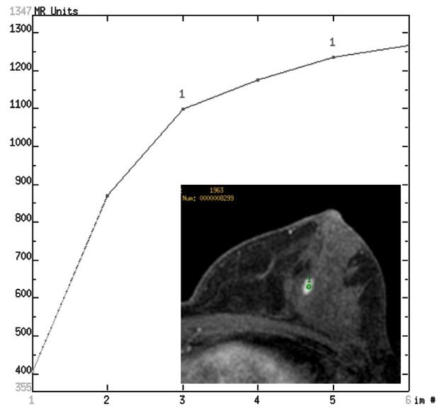
enhancement curves • Pseudoangiomatous stromal hyperplasia (MRI) - Ganzer Fall bei Radiopaedia
Breast MRI
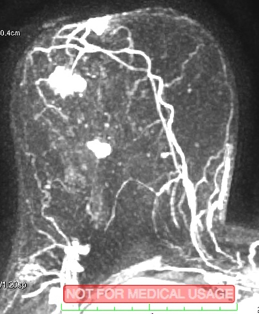
enhancement curves • High grade ductal carcinoma in situ: MRI findings - Ganzer Fall bei Radiopaedia
Breast MRI
enhancement curves • Phyllodes tumor - Ganzer Fall bei Radiopaedia
Fibroadenoma
(breast) • Breast fibroadenoma - Ganzer Fall bei Radiopaedia
Angiosarcoma
• Primary breast angiosarcoma (MRI) - Ganzer Fall bei Radiopaedia
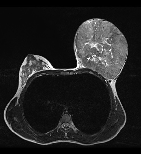
Giant
fibroadenoma • Giant fibroadenoma - Ganzer Fall bei Radiopaedia
Mucinous
carcinoma of the breast. The nodule shows a curve type I.
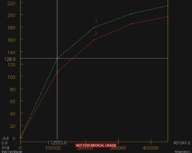
Following administration of gadolinium, there can be three possible enhancement (time intensity) kinetic curves for a lesion on breast MRI (these are also applied in other organs such as prostate MRI). These are sometimes termed the Kuhl enhancement curves.
- type I curve: progressive or persistent enhancement pattern
- typically shows a continuous increase in signal intensity throughout time
- usually considered benign with only a small proportion of (~9%) of malignant lesions having this pattern
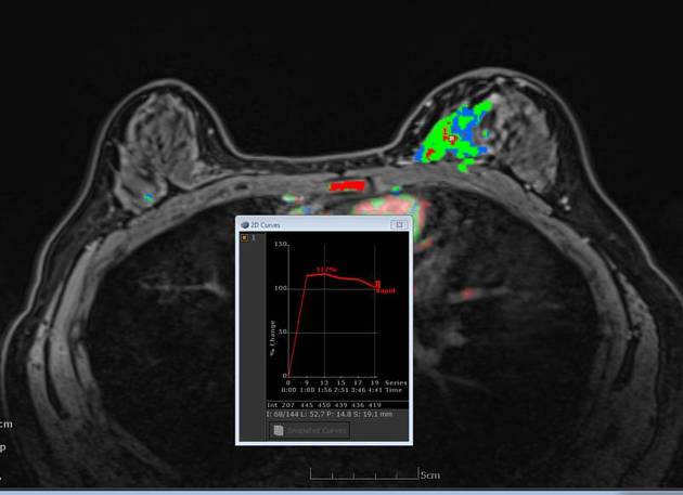
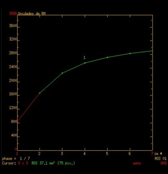
- type II curve: plateau pattern
- initial uptake followed by the plateau phase towards the latter part of the study
- considered concerning for malignancy
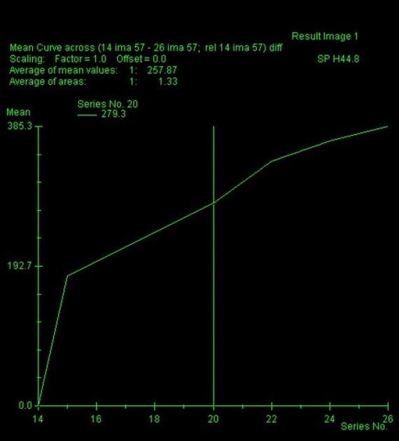
- type III curve: washout pattern
- has a relatively rapid uptake shows reduction in enhancement towards the latter part of the study
- considered strongly suggestive of malignancy
Siehe auch:
und weiter:


 Assoziationen und Differentialdiagnosen zu breast MRI enhancement curves: