Candida esophagitis

Candida esophagitis is the most common cause of infectious esophagitis that commonly affects immunocompromised patients. On imaging, it is characterized by irregular plaque-like lesions separated by normal mucosa and small (<1 cm) ulcers, which are assessed on esophagogram studies.
Epidemiology
It occurs as an opportunistic infection in immunocompromised patients, particularly those with AIDS, but it can also result from local esophageal stasis caused by severe motility disorders such as scleroderma and achalasia. Only 50% of patients have simultaneous oral thrush.
Clinical presentation
It is characterized by odynophagia in immunocompromised patients, particularly patients with AIDS.
Radiographic features
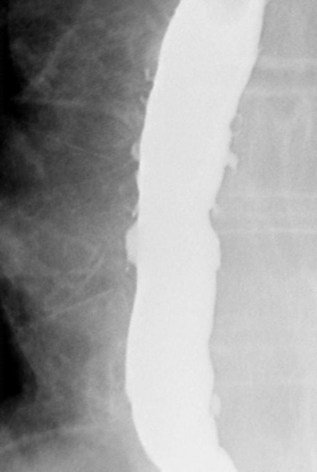
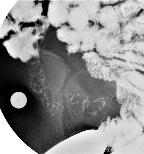
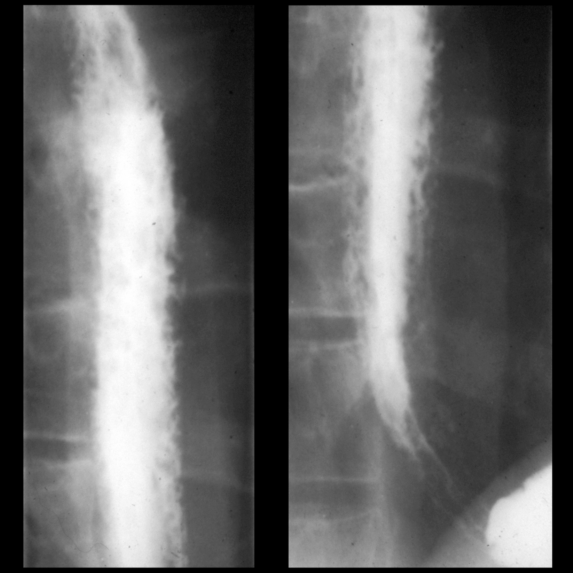
Esophagogram
On double contrast studies, it manifests as discrete longitudinally oriented linear or irregular plaque-like lesions separated by normal mucosa and small (<1 cm) punctuate, round, or oval ulcers.
In recent years a much more fulminant form has been encountered in patients with AIDS, who may present with grossly irregular or "shaggy" esophagus caused by innumerable coalescent pseudomembranes and plaques with trapping of barium between them. A cobblestone appearance may be visible.
Patients with scleroderma or achalasia may develop a "foamy" esophagus.
Differential diagnosis
Imaging differential considerations include:
- glycogenic acanthosis
- a condition affecting elderly people which result from the accumulation of glycogen in squamous epithelial cells lining the esophagus
- multiple small nodules and plaques measuring 2-10 mm in size, and of the same color of normal mucosa
- on esophagogram, it mimics candida esophagitis although its nodules have a more rounded appearance as opposed to more linear appearing plaques in moniliasis
- cytomegalovirus esophagitis
- causes ulcerations that may be up to 2 cm wide
- herpes esophagitis
- HIV esophagitis
Siehe auch:
- Achalasie
- Systemische Sklerodermie
- Intramurale Pseudodivertikulose des Ösophagus
- Ösophagitis
- foamy oesophagus
- Pflastersteinrelief
und weiter:


 Assoziationen und Differentialdiagnosen zu Moniliasis: