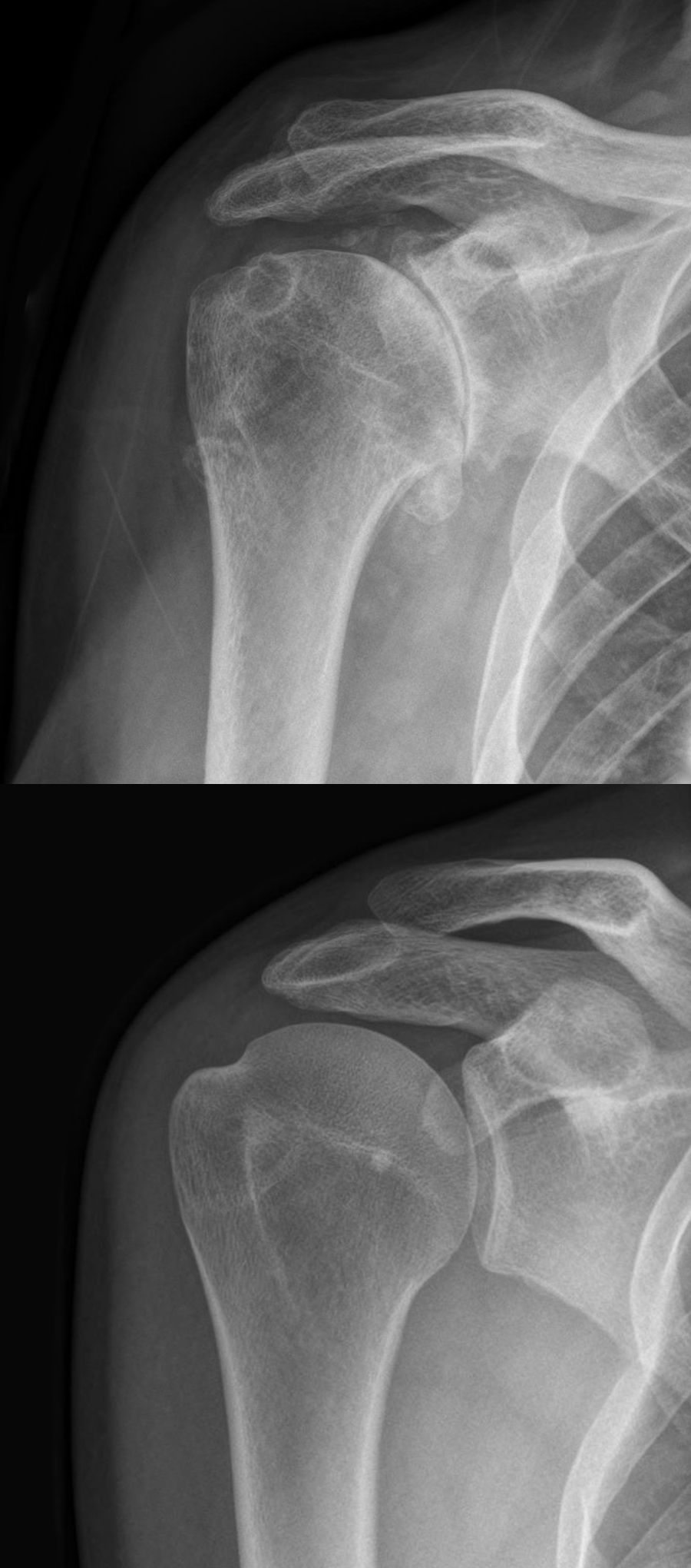
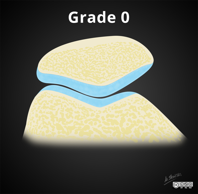
cartilage
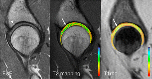
MRI for the
preoperative evaluation of femoroacetabular impingement. Sagittal PD-weighted MRI of the hip in a 30-year-old woman demonstrates mild chondral hyperintensity over the anterosuperior acetabular dome (arrowhead), with corresponding prolongation of relaxation times on T2 mapping and T1rho images (white arrows)


 Assoziationen und Differentialdiagnosen zu Knorpelbildgebung: