cavum vergae cyst
Cavum vergae cysts are rare lesions which are usually asymptomatic. It should not be confused with cavum septum pellucidum et vergae which is common. Although there is no actual pathological distinction between a run-of-the-mill cavum vergae and a cavum vergae cyst, the later is sometimes used to denote a transverse diameter of more than 1 cm or when the outer margins are convex.
Epidemiology
Cavum vergae cysts are rare congenital lesions, gradually enlarging with time. As they are often asymptomatic the diagnosis may not be made until adulthood.
Clinical presentation
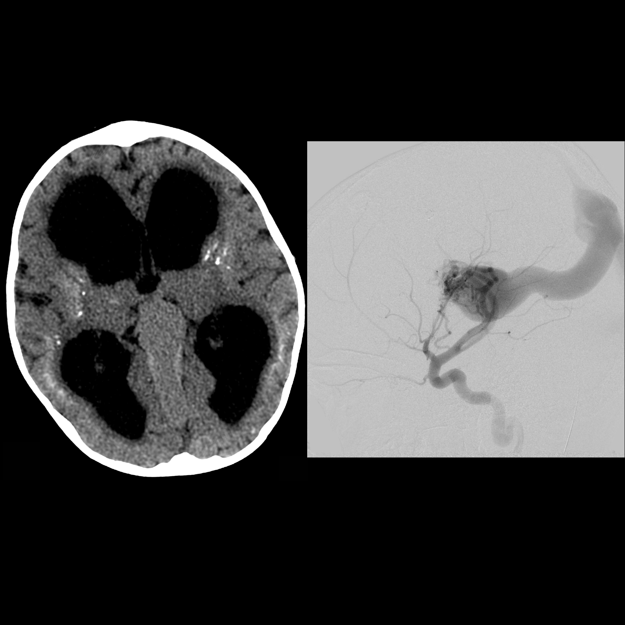
Usually asymptomatic. Occasionally the cyst is large enough to compress the midbrain tectum and aqueduct and result in obstructive hydrocephalus. Sometimes the cyst may be large enough to impair the normal drainage of the occipital and temporal horns of the lateral ventricles. In either case, presentation is with signs and symptoms of raised intracranial pressure (morning headaches, worse on stooping etc..)
Radiographic features
Ultrasound (in the neonatal or antenatal period), CT and MRI are all able to demonstrate cavum vergae cysts. Features consist of a cystic lesion located in the midline, which is located posterior to the foramen of Monro, below the posterior body and splenium of the corpus callosum and above the fornices (which it often splays).
Treatment and prognosis
In most cases, patients are entirely asymptomatic and as such, no treatment is required. If hydrocephalus is present then surgical options include:
Differential diagnosis
- cavum septum pellucidum et vergae
- cavum velum interpostium
- interhemispheric arachnoid cyst
- pineal cyst
- vein of galen aneurysm
- only a consideration on ultrasound
- color duplex scanning easily demonstrates flow
Siehe auch:


 Assoziationen und Differentialdiagnosen zu cavum vergae cyst: