Cisterna chyli


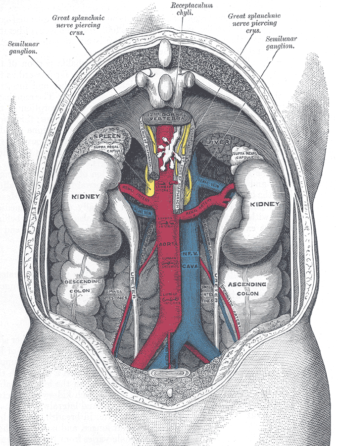

The cisterna chyli (CC) (plural cisternae chyli), also known as the receptaculum chyli, is a normal anatomical structure in the lymphatic system. It is seen as a saccular area of dilatation in the lymphatic channels that are located in the retrocrural space, usually to the immediate right of the origin of the abdominal aorta.
Gross anatomy
Location
The cisterna chyli is located at the level of L1 and L2 vertebral bodies immediately right to the aorta behind the right crus of the diaphragm.
Origin
It is an elongated, sac-like structure formed by the junction of a variable number of lumbar, intestinal, liver and descending intercostal lymphatic trunks. It extends 5-7 cm in the caudocephalad axis, but the size varies greatly with patient position.
Termination
The upper end of the cisterna chyli continues as the thoracic duct which ascends in the posterior mediastinum to empty into the left subclavian vein.
Drainage
Receives lymph from the abdominal viscera as well as the abdominal wall (below the level of the umbilicus), non-alimentary viscera and lower extremities.
Relations
- anterior: right crus of the diaphragm
- posterior: L1 and L2 vertebral bodies
- left lateral: abdominal aorta
- right lateral: azygos vein
Radiographic features
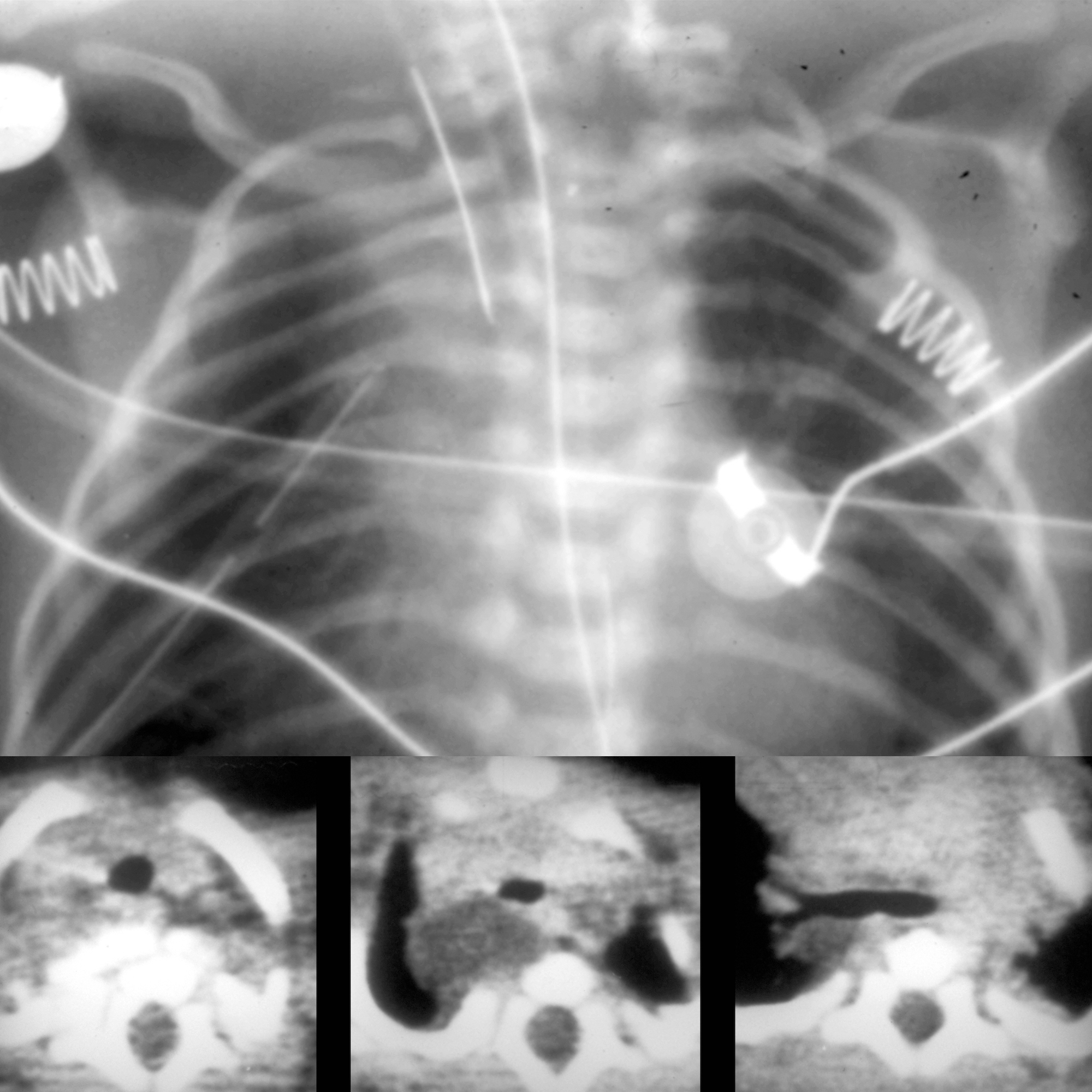
CT
The cisterna chyli can be identified as a rounded-to-elliptical retrocrural structure with an average attenuation of 4 HU. There is no enhancement following intravenous contrast administration.
MRI
The signal intensity characteristics of the cisterna chyli on MRI are the same as those for static or slow-moving fluids with high signal intensity on fluid-sensitive MR sequences .
Differential diagnosis
On CT consider:
- enlarged retrocrural lymph node
- small neurenteric cyst
Siehe auch:
und weiter:


 Assoziationen und Differentialdiagnosen zu Cisterna chyli: