epitheloides Hämangiom des Knochens
Epithelioid hemangiomas of bone are benign intraosseous vascular neoplasms of epithelioid morphology which show locally aggressive behavior.
Epidemiology
Epithelioid hemangiomas of bone are uncommon tumors with an unknown incidence. They have been observed in all age groups, with men being slightly more frequently affected .
Clinical presentation
The most common complaint is pain. They are rarely found incidentally .
Pathology
Epithelioid hemangiomas of bone are expansile vascular tumors with a lobular architecture sometimes showing cortical erosion and extension of the soft tissues .
Location
About 18-25% of epithelioid hemangiomas of bone have been found in a multifocal regional distribution involving the following bones. The metaphysis or diaphysis is usually affected. There might be epiphysial involvement in pediatric patients.
- long tubular bones (≈40%)
- short tubular bones (feet > hands)
- flat bones
- vertebrae
Macroscopic appearance
Macroscopically epithelioid hemangiomas of bone are usually solid, soft, nodular, well-defined tumors of red color due to hemorrhagic components which replace the bone marrow cavity .
Microscopic appearance
Microscopic features epithelioid hemangiomas include the following :
- lobular architecture with large epithelioid, endothelial cells with eosinophilic cytoplasm
- arteriole-like vessels in the periphery of the lesions
- infrequent not atypical mitoses
- possibly small foci of necrosis
- stroma consisting of connective tissue sometimes with inflammatory infiltrates
- sometimes foci of intralesional hemorrhage
Immunohistochemistry
Immunohistochemistry stains are usually positive for endothelial markers as CD31, CD34, FLI1, ERG or factor VIII-related antigen and might be positive for keratin or EMA.
Genetics
The pathogenesis of epithelioid hemangioma of bone involves a fusion including the FOS gene at 14q24.3 or alternatively the FOSB gene at 19q13.32 .
Radiographic features
General imaging features of epithelioid hemangioma of bone are the following :
- lobular architecture with vascular formations
- well-defined margins
- intralesional septae
- expansile growth
- occasional cortical erosions and soft tissue extension
Plain radiograph
On plain radiographs epithelioid hemangioma will usually display the following characteristics :
- expansile radiolucent, lytic or cystic-appearing lesions
- narrow transition zone
- endosteal scalloping
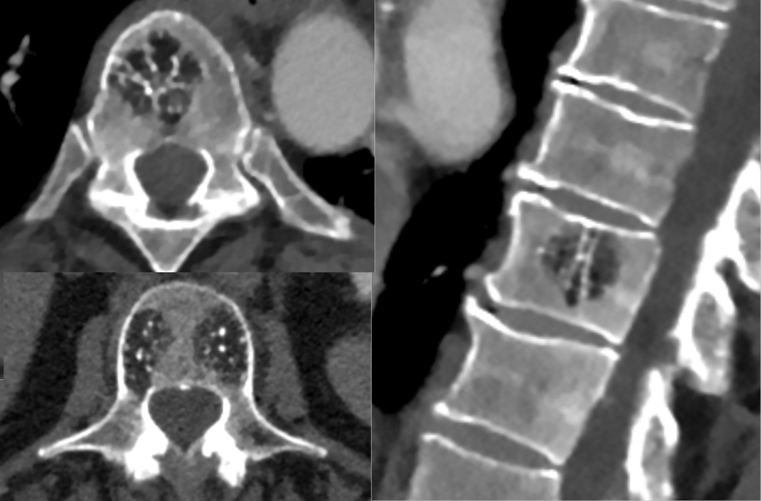
CT
Similar to plain radiographs CT will show an expansile lytic or cystic lesions with an inner soft tissue component usually slightly lower than muscle density and no calcified or osteoid matrix .
MRI
MRI will usually show lobulated lesions with surrounding bone marrow edema and intralesional flow voids, otherwise, features of epitheloid hemangioma are non-specific .
- T1: low to intermediate signal intensity
- T2: heterogeneous, intermediate to high signal intensity
- T1 C+ (Gd): avid homogeneous enhancement
Radiology report
The radiological report should include a description of the following:
- form, location and size
- tumor margins and transition zone
- cortical erosion, cortical breakthrough
- soft tissue component
Treatment and prognosis
The tumor management usually includes curettage or cryosurgery, rarely en bloc resection . In inaccessible locations, the tumor has been irradiated .
The prognosis is good with local recurrences in about 10%. Regional lymph node involvement has been observed, however, it is unclear whether this reflects metastatic deposits or multifocal disease .
Differential diagnosis
Conditions or tumors which can mimic the presentation and/or the appearance of epithelioid hemangiomas include :
- angiosarcoma
- epitheloid hemangioendothelioma
- Langerhans cell histiocytosis
- enchondromatosis: in small tubular bones
- chondromyxoid fibroma
- giant cell tumor
- aneurysmal bone cyst
See also
Siehe auch:


 Assoziationen und Differentialdiagnosen zu epitheloides Hämangiom des Knochens: