Keilbeinhöhle


The sphenoid sinus is the most posterior paranasal sinus.
Summary
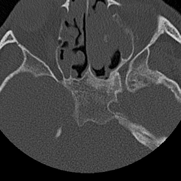
- location: the central body of the sphenoid bone anteroinferior to the sella turcica
- blood supply: posterior ethmoidal and sphenopalatine arteries
- innervation: posterior ethmoidal nerve and the orbital branch of the pterygopalatine ganglion
Gross anatomy
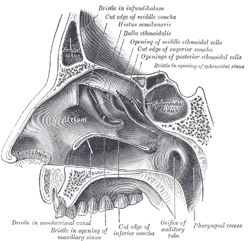
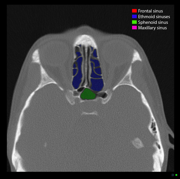
The sphenoid sinuses are paired spaces formed within the body of the sphenoid bone, communicating with the roof of the nasal cavity via the sphenoethmoidal recess in its anterior wall. The two sinuses are separated by a septum which may or may not be in the midline. It usually lies anteroinferior to the sella.
A large sinus can show a number of ridges and depressions related to closely adjacent structures. These can include the pituitary gland, optic nerve, and internal carotid artery.
Relations
- superiorly: cavernous sinus, sella turcica, and its contents
- inferiorly: nasal cavities
- anteriorly: nasal cavities, posterior ethmoid air cells
- posteriorly: contents of the middle cranial fossa
- laterally: cavernous sinus, cranial cavity
Types of pneumatization
- conchal
- lateral recess
- presellar
- sellar (which can be incomplete or complete)
Arterial supply
- posterior ethmoidal artery
- nasal branches of the sphenopalatine artery
Venous drainage
- superior ophthalmic veins via posterior ethmoidal veins
Lymphatic drainage
Lymph drainage occurs via afferent vessels leading into the retropharyngeal nodes
Innervation
- parasympathetic: orbital branches of the pterygopalatine ganglion
- sensory: posterior ethmoidal nerve from the nasociliary nerve, a branch of the ophthalmic division of the trigeminal nerve
Variant anatomy
With respect to optic nerve relationship:
- type 1: adjacent to sphenoid sinus
- type 2: indenting the sinus
- type 3: traversing the sinus
- type 4: adjacent to posterior ethmoid sinus
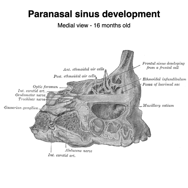
Development
Pneumatization starts at around 2 years of age and it develops more slowly than the other paranasal sinuses.
Practical points
It is important to look for and report the type of pneumatization of this sinus and to report the relationship with adjacent neurovascular structures, especially dehiscence.
See also
Siehe auch:
und weiter:


 Assoziationen und Differentialdiagnosen zu Sinus sphenoidalis: