lipomyelomeningocele
Lipomyelomeningoceles are one of the forms of closed spinal dysraphism. They usually present as a subcutaneous fatty mass just above the intergluteal cleft. However, some lipomyelomeningoceles may occur at other locations along the spinal canal.
Clinical presentation
Lipomyelomeningoceles may be clinically detectable as a subcutaneous fatty mass above the intergluteal crease.
Pathology
These lesions are formed mainly due to a defect in primary neurulation in which mesenchymal tissue enters into neural placode and forms lipomatous tissue.
Radiographic features
Plain radiograph
It shows a non-specific splaying / non-fusion of the posterior elements of the spine, which usually cannot be differentiated from the other spinal dysraphisms.
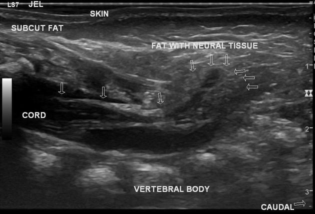
Ultrasound
Ultrasound will show the vertebral arch defector defect associated with an elongated spinal cord with the placode attached to an intrathecal echogenic fatty mass that protrudes in the dorsal subcutaneous tissue (see case 2).
CT
- spina bifida
- focal enlargement of the spinal canal at the placode level
- hypodense fat-attenuated mass in continuity with the subcutaneous fat planes
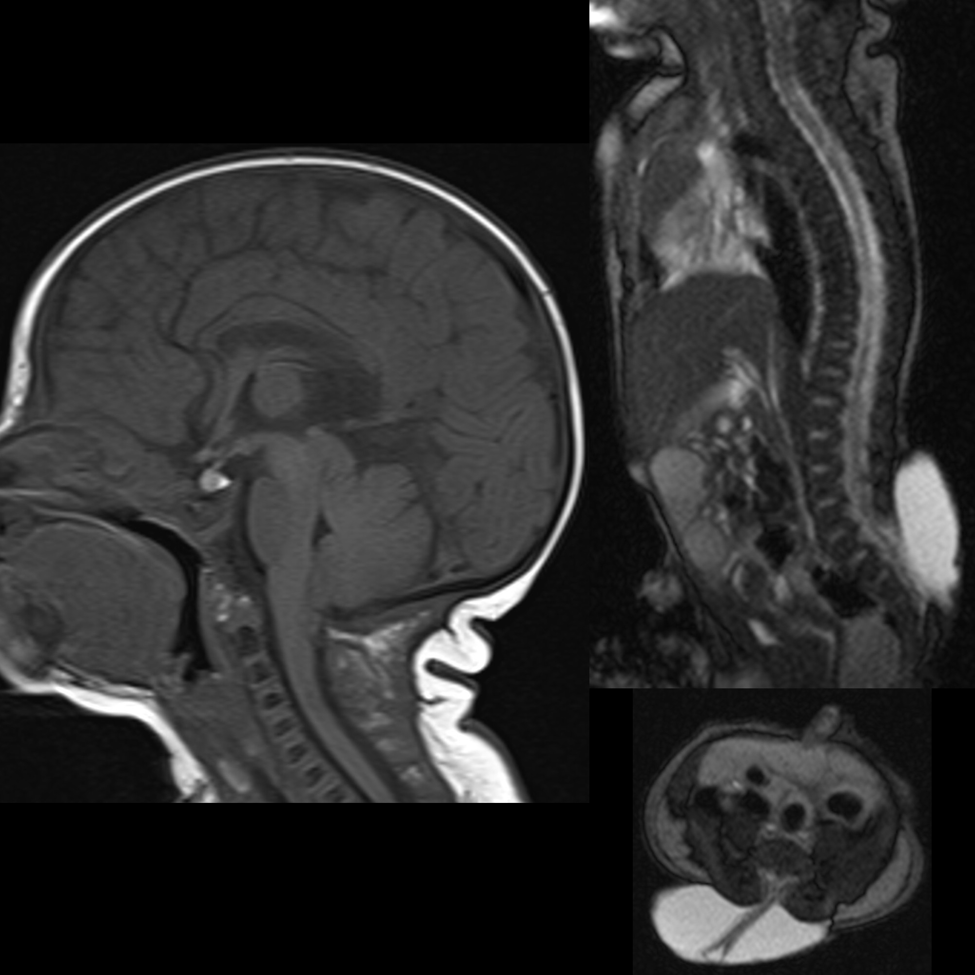
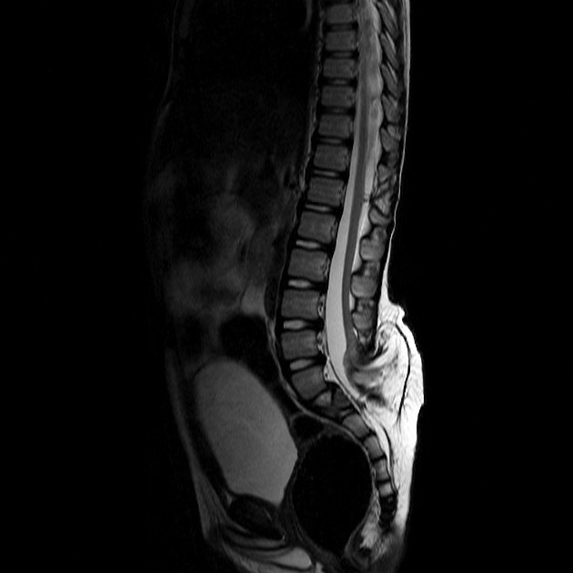
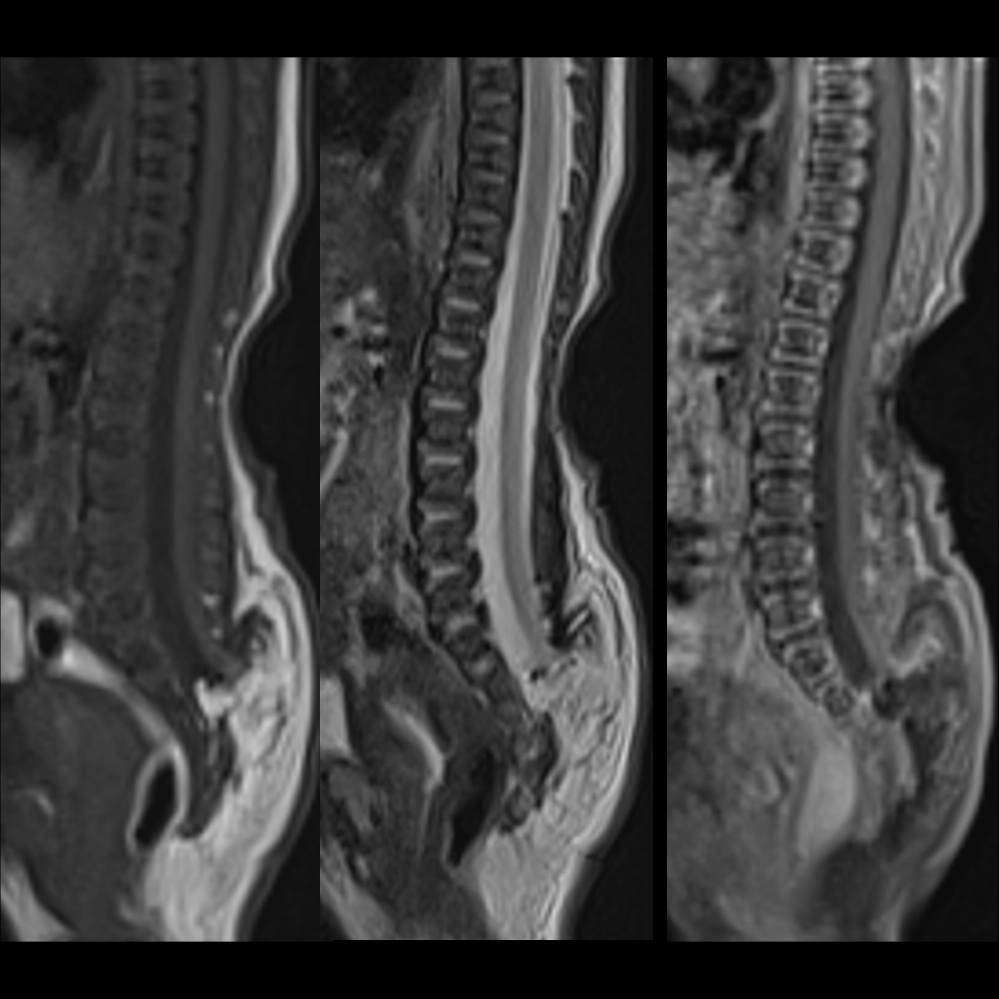
MRI
- typically seen as a spinal defect with lipomatous tissue, covered with skin
- neural placode-lipoma interface lies outside the spinal canal due to enlargement of subarachnoid space
- a low-lying cord is usually present
Differential diagnosis
- lipomyelocele: placode-lipoma interface lies within the spinal canal
See also
Siehe auch:


 Assoziationen und Differentialdiagnosen zu lipomyelomeningocele: