occipital lobe


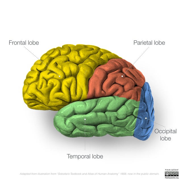
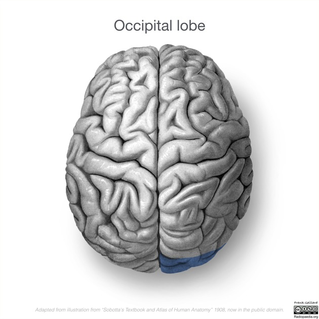
The occipital lobe is the smallest of the four lobes of the brain. It sits posterior to the temporal lobe and parietal lobes, underlying the occipital bone and overlying the tentorium cerebelli. Its most notable functional component is the primary and secondary visual cortex.
Gross anatomy
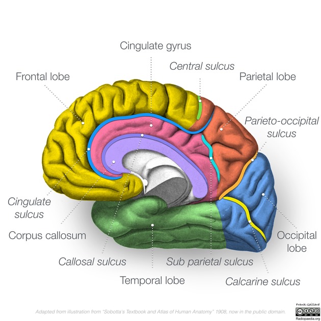
The occipital lobe is the smallest lobe accounting for only 18% of the total neocortical volume. The boundaries of the occipital lobe have been arbitrarily defined, giving it a triangular shape . It is separated from the parietal and temporal lobes on the medial surface by the parieto-occipital sulcus and on the lateral side by the lateral parietotemporal line, a near vertical imaginary line extending from the preoccipital notch of the temporal lobe superiorly to the parieto-occipital sulcus. Medially, it is confined by the medial longitudinal fissure which divides both cerebral hemispheres. The tentorium cerebelli forms its inferior border and the occipital bone its posterior margin.
Functional areas
The occipital lobe is concerned with visual processing and is composed of three Brodmann areas:
- primary visual cortex (Brodmann area 17)
- secondary visual (association) cortex (Brodmann areas 18 and 19)
Sulci and gyri
The occipital lobe has a predictable medial gyral anatomy.
- medial surface (from superior to inferior)
- parieto-occipital sulcus
- cuneus
- calcarine sulcus
- site of the primary visual cortex
- lingual gyrus
- collateral sulcus
- posterior segment, extending from temporal lobe
- fusiform gyrus
The lateral surface of the occipital lobe is highly variable, reflected by the literature which is either vague or contradictory. As such, care must be taken in using specific anatomical terms as readers may have a different interpretation of the same anatomy.
The simplest description, which conforms to the sulcal pattern found in the frontal and temporal lobes, unfortunately, is only present in approximately two-thirds of cases . When present the lateral surface is divided into three roughly parallel gyri (superior, middle and inferior occipital gyri) by two transverse occipital sulci (referred to as superior and inferior when both are present).
Relations
- anterior: parietal lobe and temporal lobe
- posterior: occipital bone
- superior: parietal lobe and parietal bone
- inferior: tentorium cerebelli
- medial: transverse sinuses, torcula
Blood supply
- branches of the posterior cerebral artery (PCA)
- parieto-occipital artery
- calcarine artery
- posterior temporal artery
- common temporal artery/ temporo-occipital artery/ lateral occipital artery
- lingual gyrus artery
- not found in all individuals
Neurological deficits
The following neurological deficits occur with unilateral or bilateral lesions of the occipital lobes :
- deficits arising from unilateral lesions involving the dominant hemisphere:
- hemianopia: retrochiasmal lesion (lesions involving the optic tract, thalamic lateral geniculate nucleus, occipital lobe)
- color dysnomia: interruption of fibers streaming from the occipital cortex to the Wernicke's area
- Anton syndrome: those who suffer cortical blindness but affirm quite adamantly that they are able to see
- irritative lesions involving either lobe can give rise to the following:
- visual hallucinations (e.g. seeing flashes of light)
Siehe auch:


 Assoziationen und Differentialdiagnosen zu Okzipitallappen: