Os odontoideum

 nicht verwechseln mit: Persistierendes Ossiculum terminale
Os odontoideum (plural: ossa odontoidea) is an anatomic variant of the odontoid process of C2 and needs to be differentiated from persistent ossiculum terminale and from a type 2 odontoid fracture. It can be associated with atlantoaxial instability.
Although it was originally thought to be a congenital lesion due to a failure of the center of ossification of the dens to fuse with the body of C2, it may actually represent an unremembered and/or unrecognised fracture through the dens's growth plate before the age of 5 or 6. There may be associated instability and chronic symptoms .
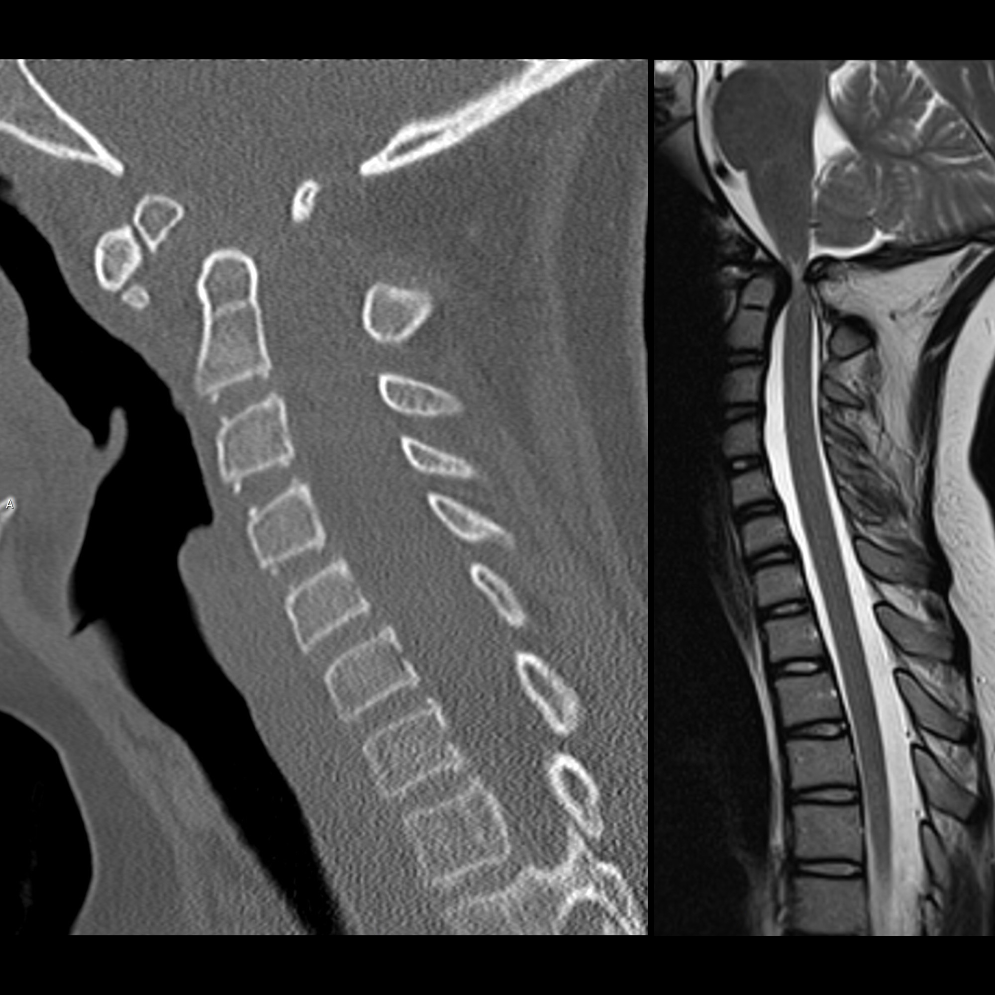
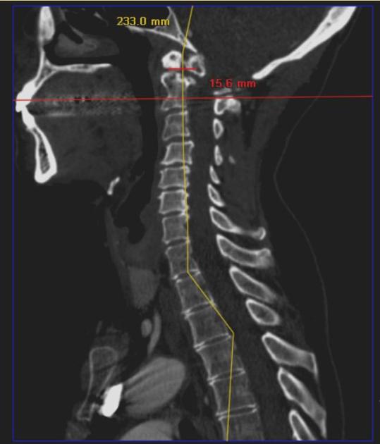
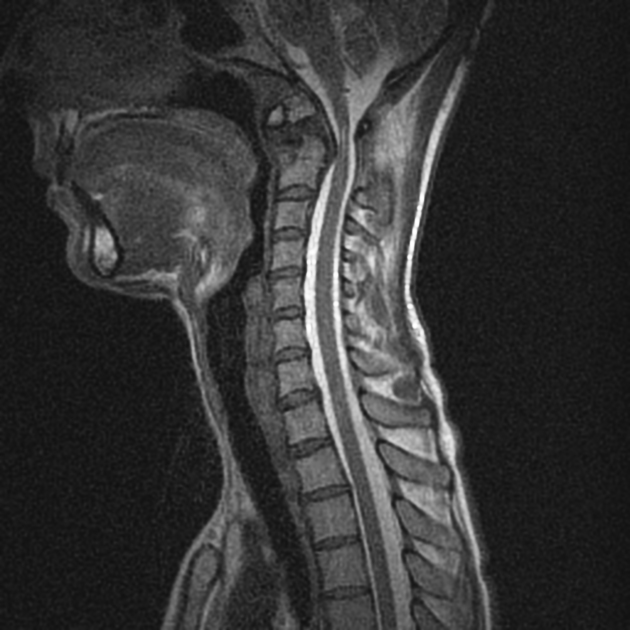
The level of mobility is below the transverse band of the cruciform ligament and therefore results in abnormal mobility of the dens with respect to C2.
Subtypes
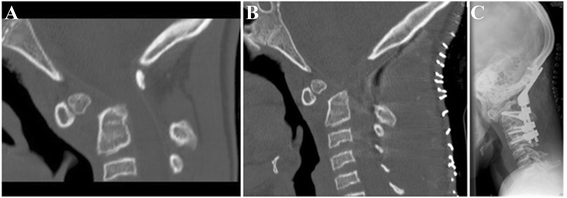
An os odontoideum can be divided into two main types :
- orthotopic: normal position with a wide gap between C2 and os odontoideum
- dystopic: displaced (dystopic os odontoideum has also been termed the "os avis" by some)
Associations
Often discovered incidentally on its own. Associated syndromes include:
It may be seen in association with another adjacent anatomic variant, the third condyle .
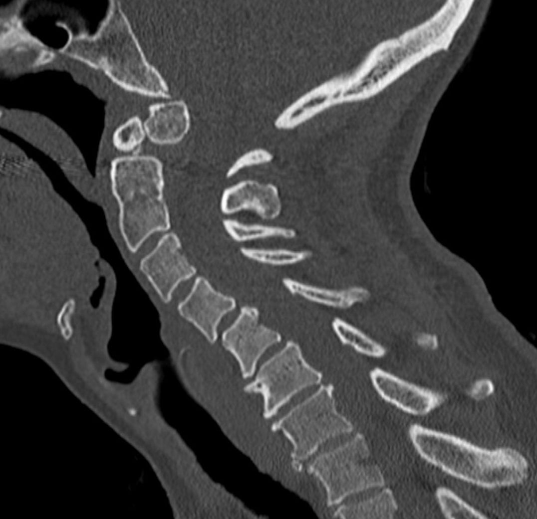
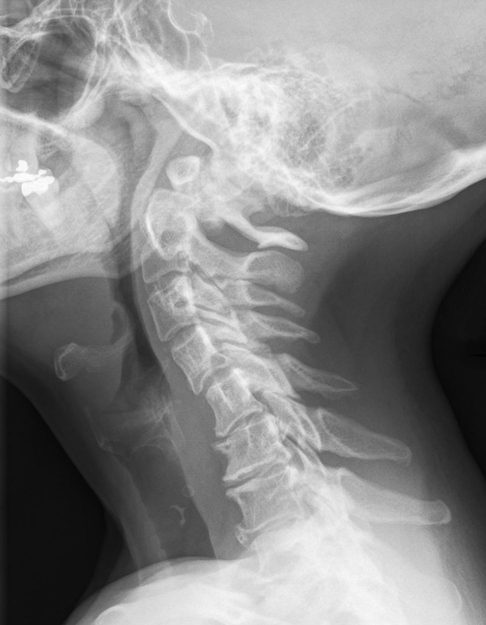
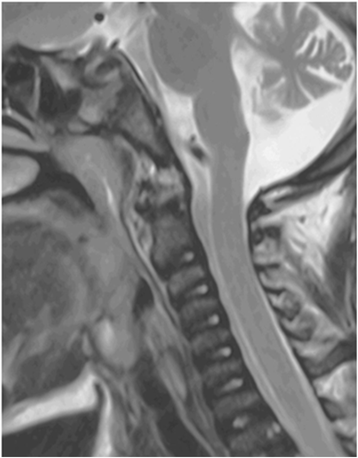
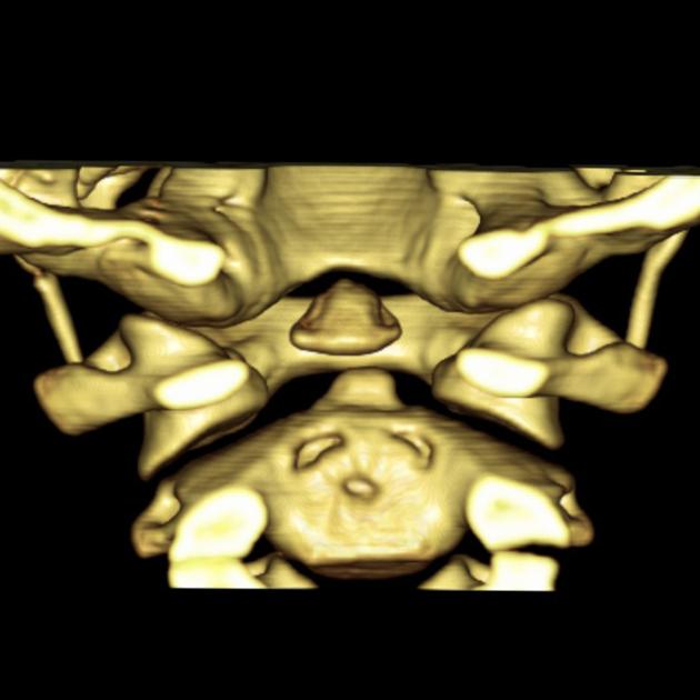
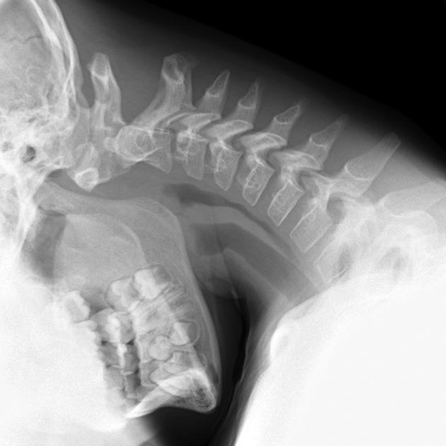
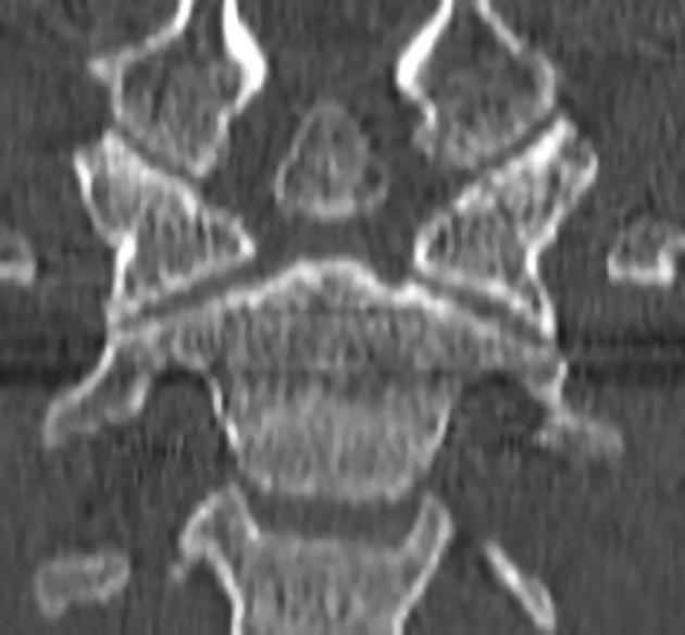
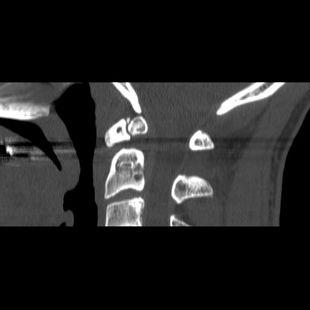
Radiographic features
- smooth, well-corticated ossicle at the superior ossicle of a hypoplastic dens
- around half the size of a normal dens
- associated with hypertrophied and rounded anterior arch of the atlas
History and etymology
The Italian anatomist and neuroscientist Carlo Giacomini (1840-1898), first described the os odontoideum in humans in 1886. He is now better remembered for the band of Giacomini (in the hippocampal formation) and vein of Giacomini .
Differential diagnosis
Os odontoideum is one of the skeletal “don’t touch” lesions.
Siehe auch:
- Pseudarthrose Densfraktur
- Densfraktur
- Anomalien des kraniozervikalen Übergangs
- Axis Varianten
- Condylus tertius
- Persistierendes Ossiculum terminale
und weiter:


 Assoziationen und Differentialdiagnosen zu Os odontoideum:
