Pellegrini-Stieda lesion


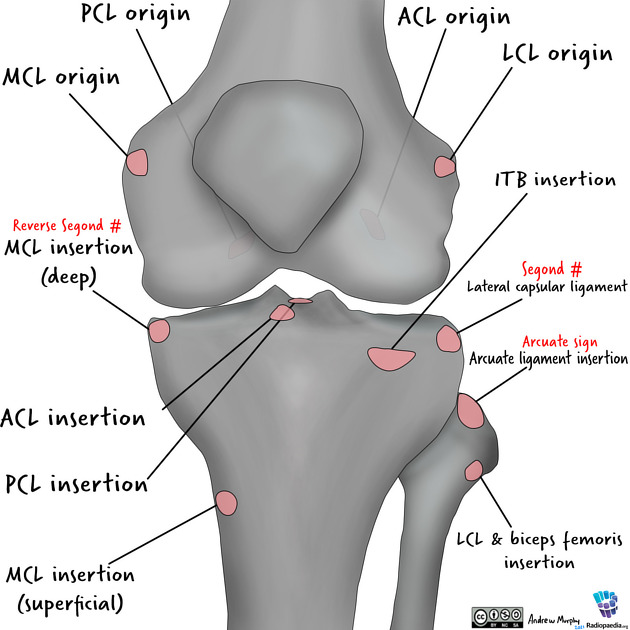
Pellegrini-Stieda lesions are ossified post-traumatic lesions at (or near) the medial femoral collateral ligament adjacent to the margin of the medial femoral condyle. One presumed mechanism of injury is a Stieda fracture (avulsion injury of the medial collateral ligament at the medial femoral condyle). Calcification usually begins to form a few weeks after the initial injury.
Clinical presentation
Most patients are asymptomatic while a small proportion will have medial knee pain (Pellegrini-Stieda syndrome).
Radiographic features
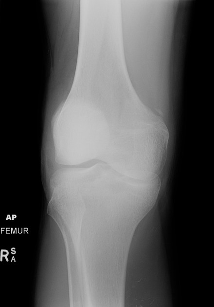
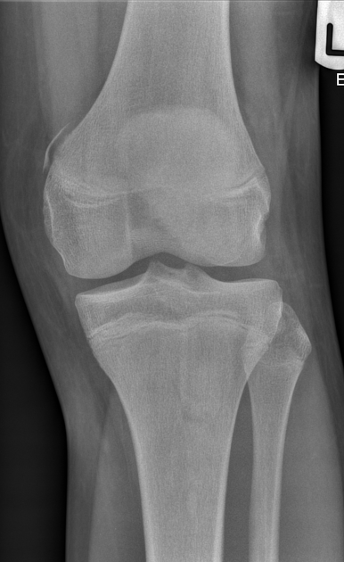
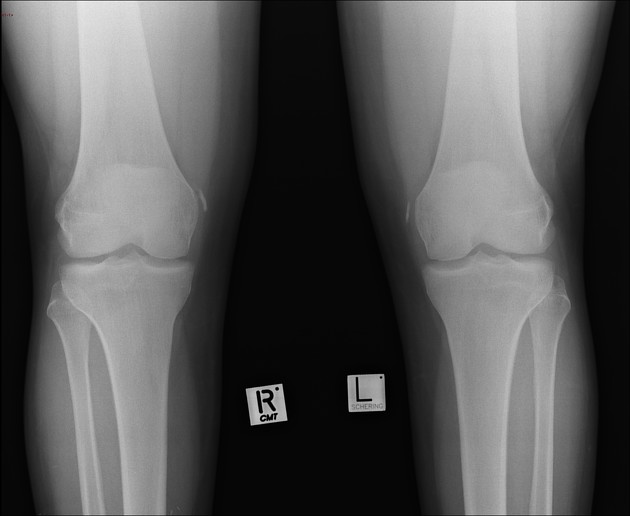
Plain radiograph
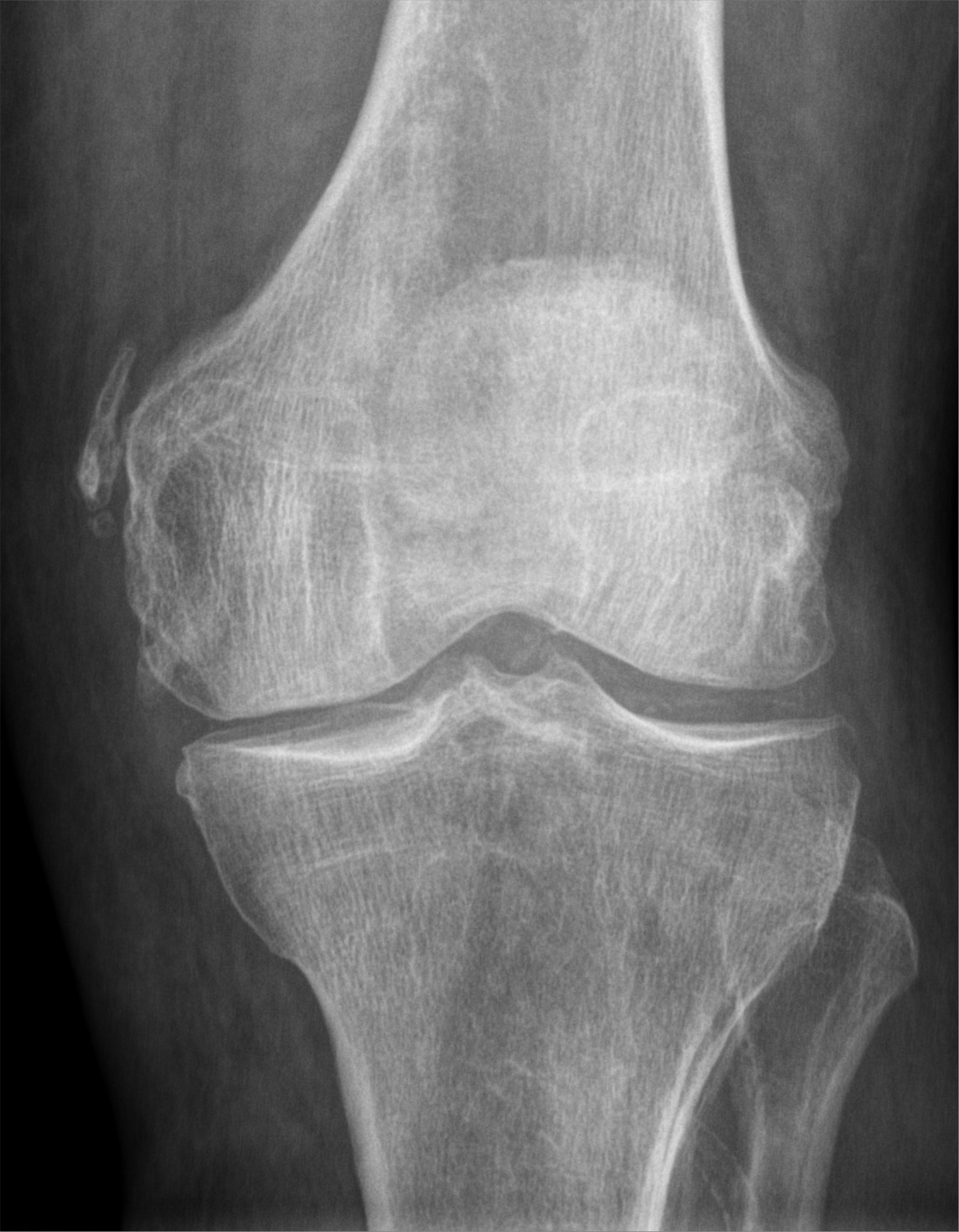
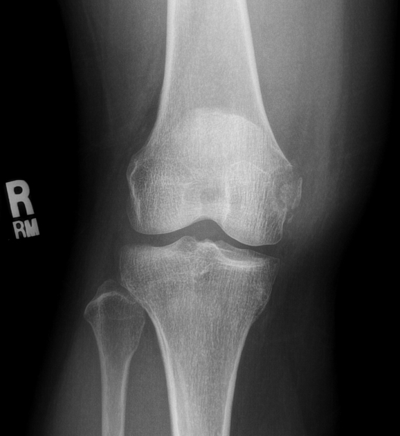
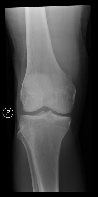
Calcification adjacent to the medial femoral condyle, often linear or curvilinear in shape and paralleling the femoral cortex.
MRI
It appears as an ossicle or enthesophyte showing bone marrow signal at the medial femoral condyle. The medial collateral ligament is usually thickened.
Treatment and prognosis
Mild and moderate cases are often conservatively managed with steroid injections and range-of-motion exercises. Surgical excision of calcifications and MCL repair is considered mainly for refractory cases .
History and etymology
It is named after Italian surgeon Augusto Pellegrini (1877-1958) and German surgeon Alfred Stieda (1869-1945).
Differential diagnosis
- tendinous calcification in reactive arthritis: often exhibits other degenerative changes
Siehe auch:
- medial collateral ligament injury grading
- Avulsionsfrakturen am Knie
- medial collateral ligament injury
- Processus Stieda
- Stieda-Pellegrini Syndrom
- lateral collateral ligament injury of the knee
und weiter:


 Assoziationen und Differentialdiagnosen zu Stieda-Pellegrini-Köhler-Schatten: