pleurale Tumoren

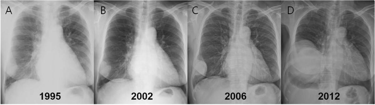
Pleuramesotheliom
im Röntgenbild: Zirkuläre Verbreiterung der Pleura links mit Pleuraerguss.

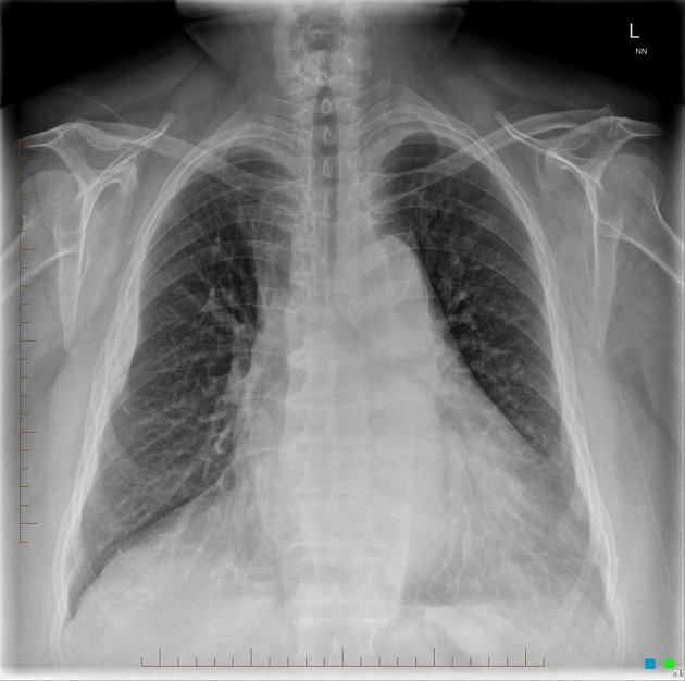
This PA chest
radiograph demonstrates an abnormal contour in the right hilar region, with visualization of the pulmonary vessels through the mass (the hilar overlay sign) indicating its posterior mediastinal location. On resection this was found to be a benign solitary fibrous tumor of the pleura.

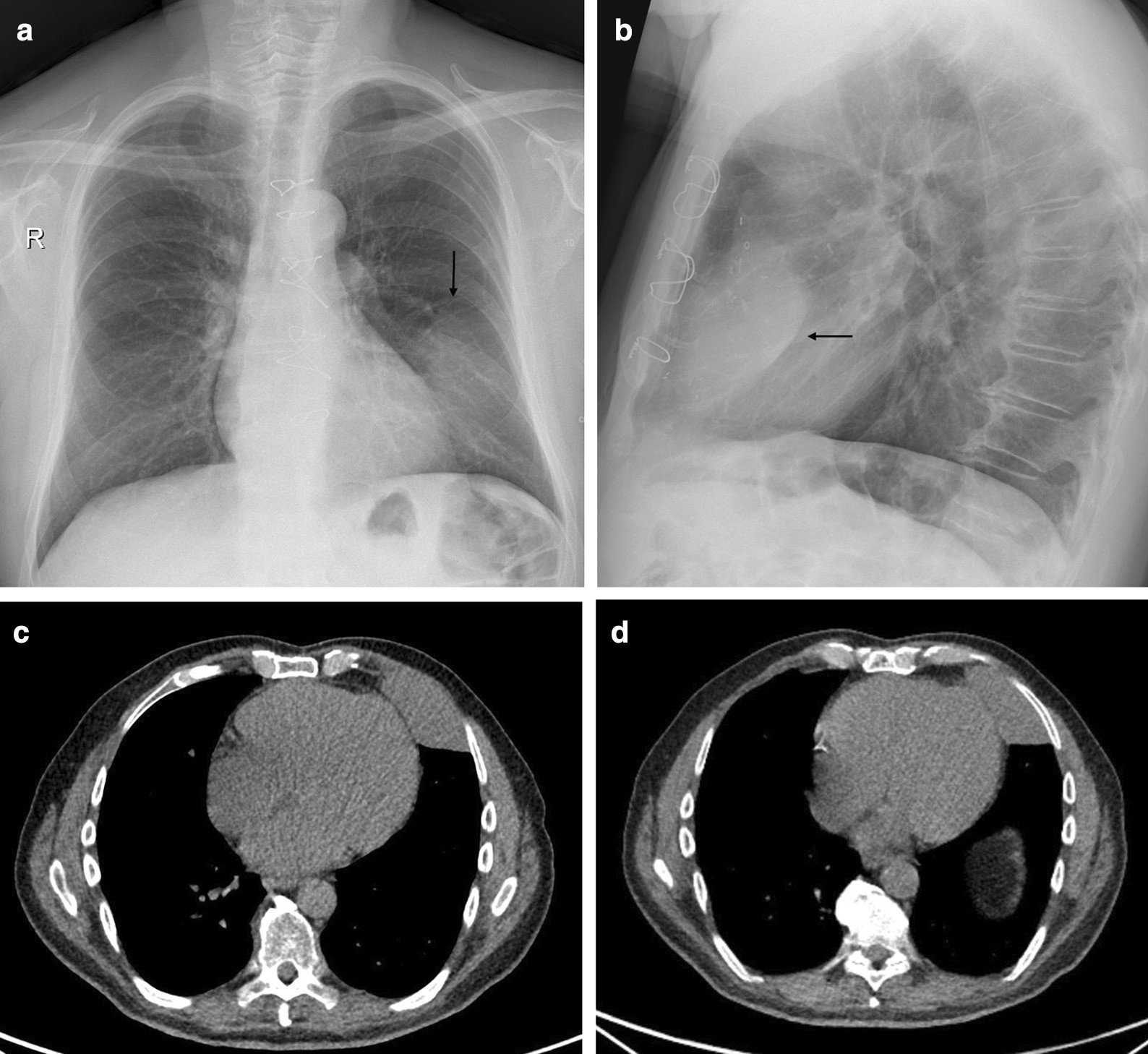
A case of
malignant solitary fibrous tumour of the mediastinal pleura. Axial chest CT image shows a well-circumscribed round enhancing solid mass located in the middle part of the mediastinum.

A case of
malignant solitary fibrous tumour of the mediastinal pleura. Coronal chest CT image shows a well-circumscribed round mild heterogeneous enhancing solid mass located in the middle part of the mediastinum, with left dislocation of the oesophagus (arrows).

A case of
malignant solitary fibrous tumour of the mediastinal pleura. Coronal chest CT image shows a well-circumscribed round enhancing solid mass located in the middle part of the mediastinum, with eccentric calcification (arrow).

Desmoplastic
round cell tumour of pleura with liver and spine metastases: Uncommon pathology with grave prognosis. Coronal reformation demonstrates multiple hypodense lesions in the lower thoracic spine (metastasis) with heterogeneously enhancing nodular thickening on LT side causing collapse of underlying lung parenchyma.


 Assoziationen und Differentialdiagnosen zu pleurale Tumoren: