Pneumomediastinum


Pneumomediastinum is the presence of extraluminal gas within the mediastinum. Gas may originate from the lungs, trachea, central bronchi, esophagus, and peritoneal cavity and track from the mediastinum to the neck or abdomen.
Terminology
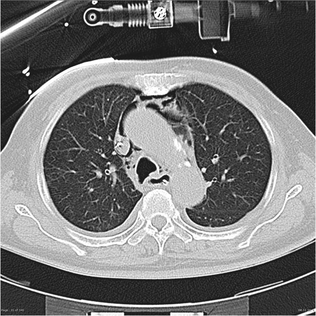
In the setting of trauma, if pneumomediastinum is visible on chest x-ray it is termed overt pneumomediastinum whereas if it is only visible on CT then it is termed occult pneumomediastinum .
Pathology
Etiology
- blunt or penetrating chest trauma
- secondary to neck, thoracic, or retroperitoneal surgery
- esophageal perforation
- Boerhaave syndrome
- endoscopic intervention
- esophageal carcinoma
- tracheobronchial perforation
- laceration
- bronchial stump dehiscence
- bronchoscopy
- tracheostomy
- laryngeal fracture
- vigorous exercise (see: pulmonary interstitial emphysema)
- childbirth (see: Hamman syndrome)
- weightlifting
- Valsalva maneuver
- asthma
- barotrauma
- diving
- ventilator: most commonly secondary to ARDS with positive pressure ventilation
- infection
- interstitial lung disease
- connective tissue disorders
- polymyositis/dermatomyositis, uncertain if related to vasculopathy or associated interstitial lung disease
- idiopathic
Although it is rare, pneumomediastinum can occur spontaneously. This is considered benign and generally affects young adult males .
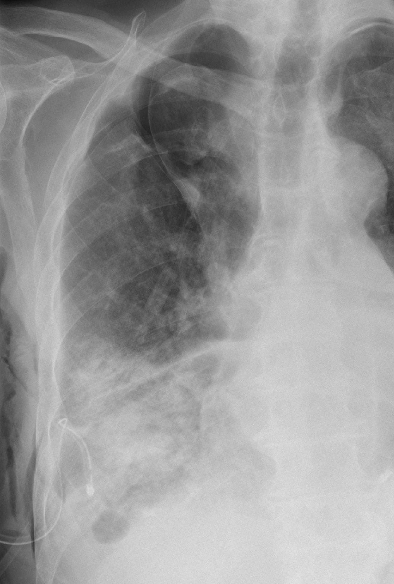
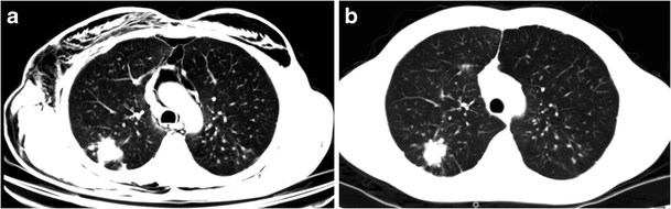
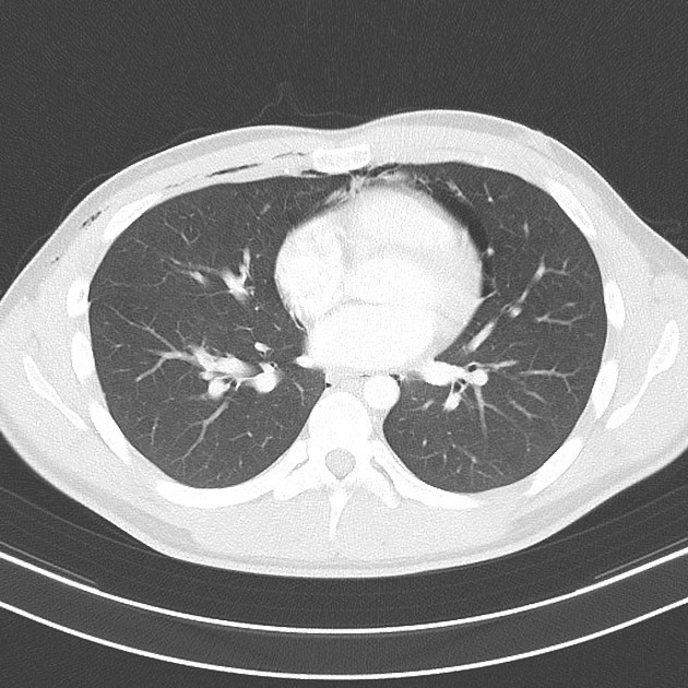
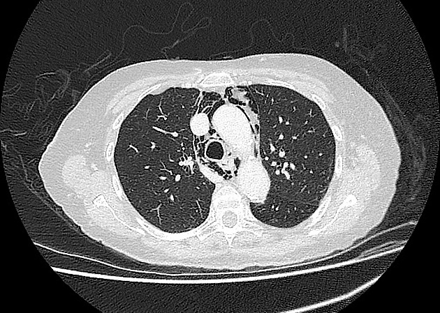
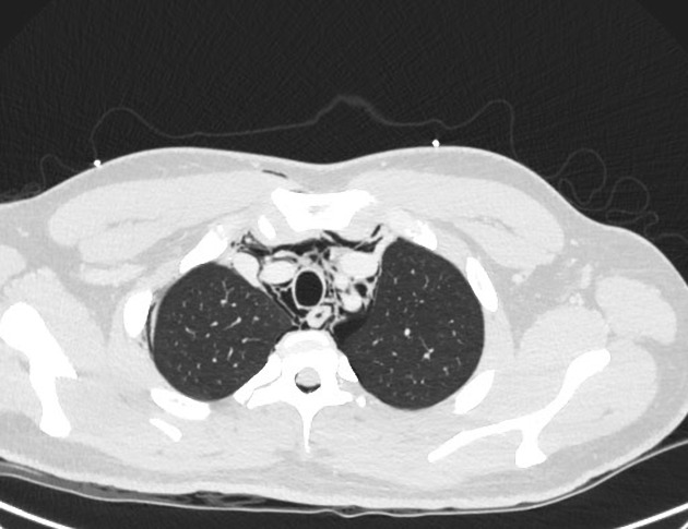
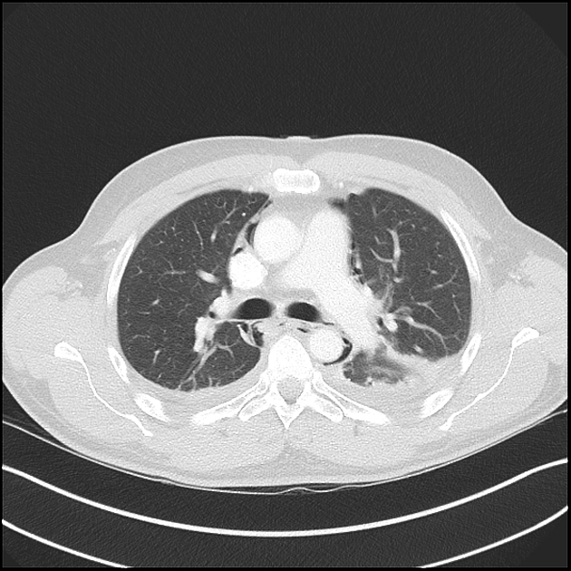
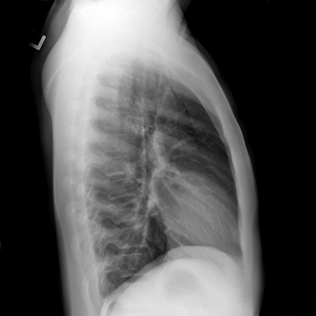
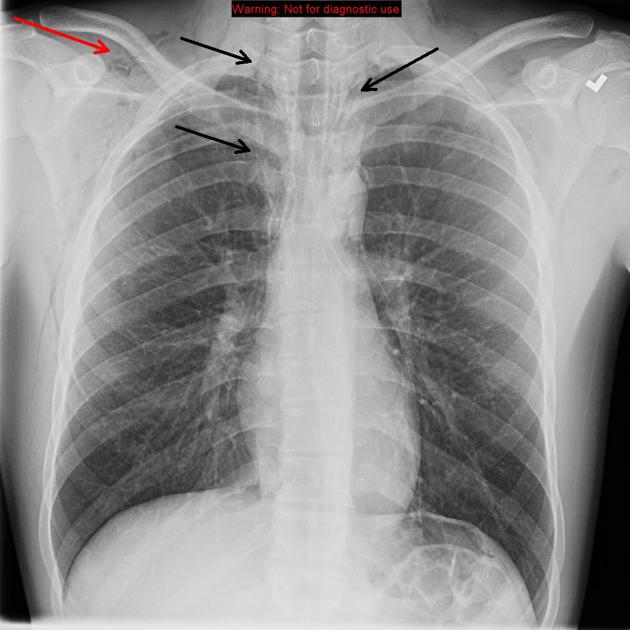
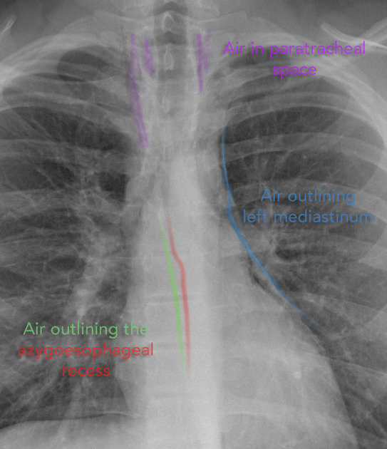
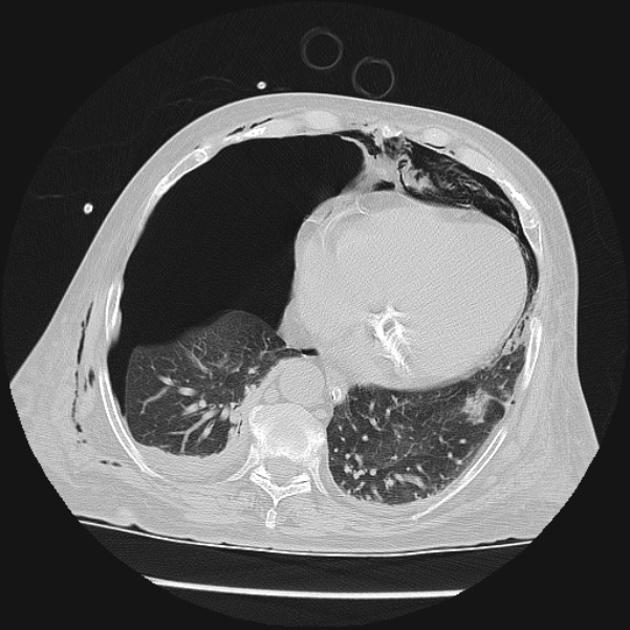
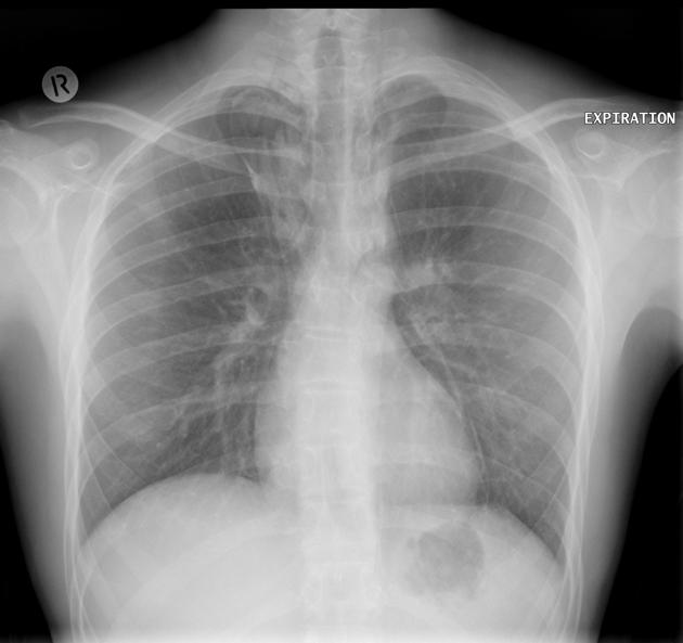
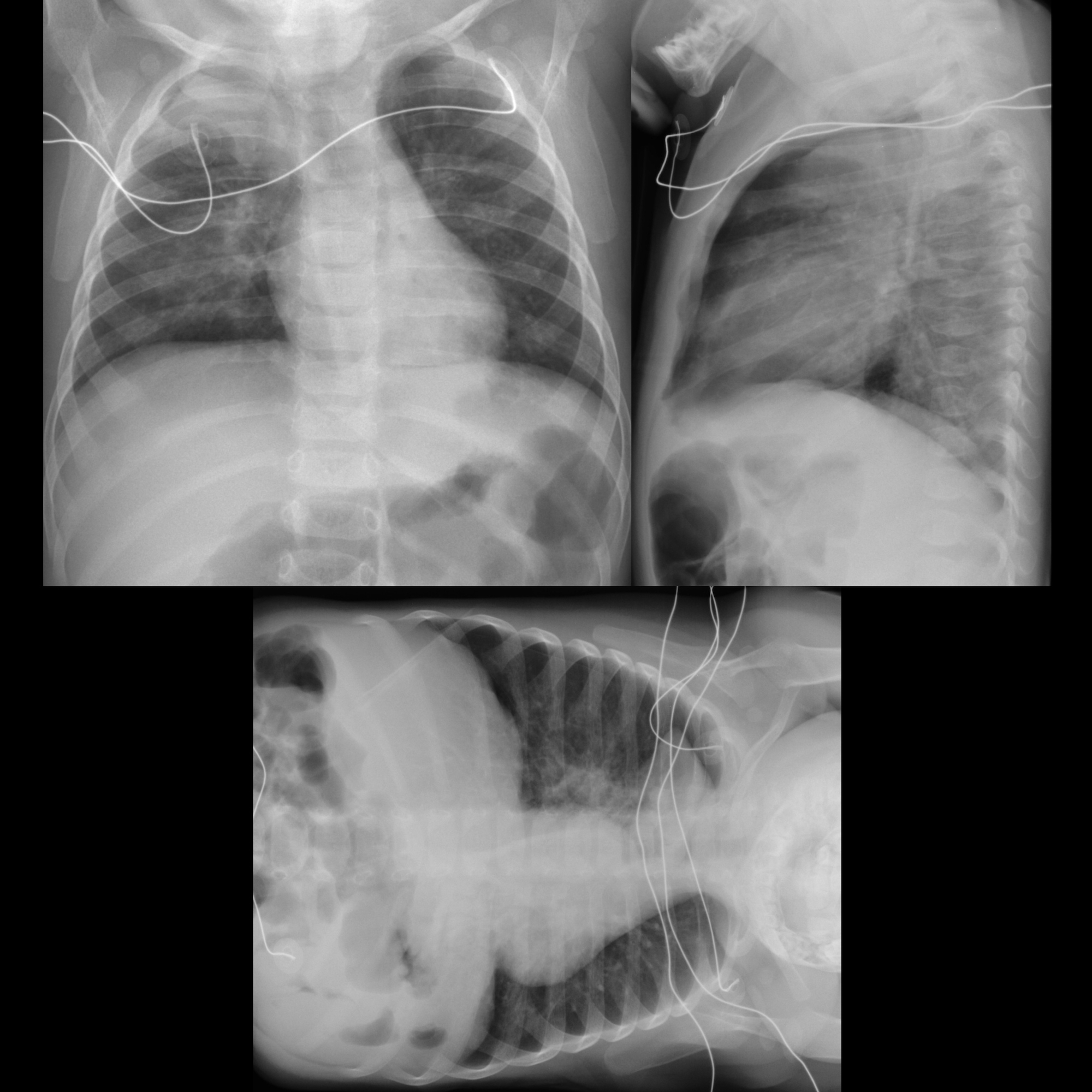
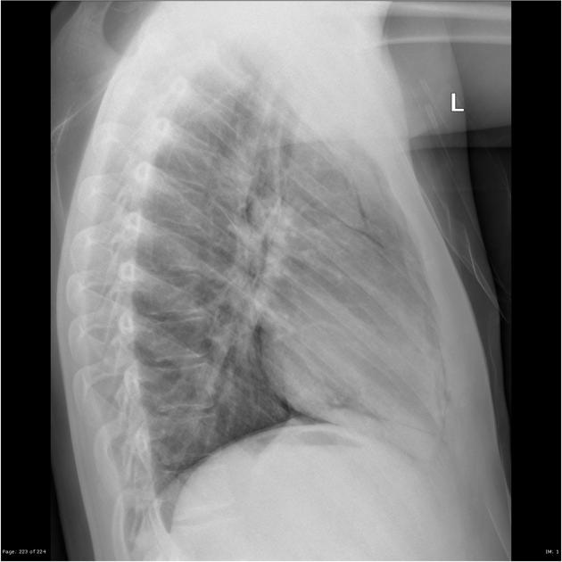
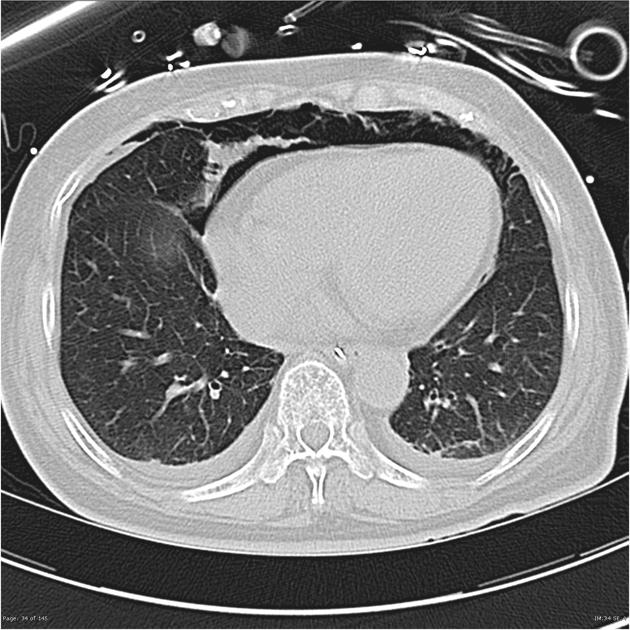
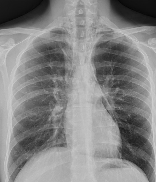
Radiographic features
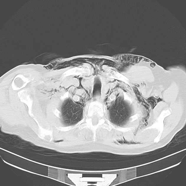
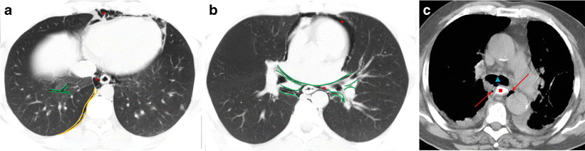
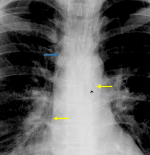
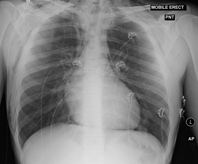
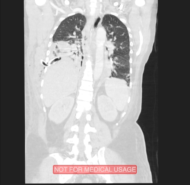
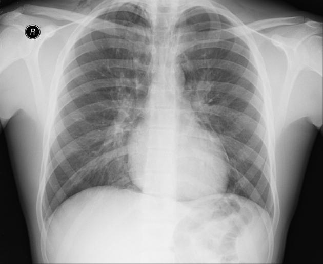
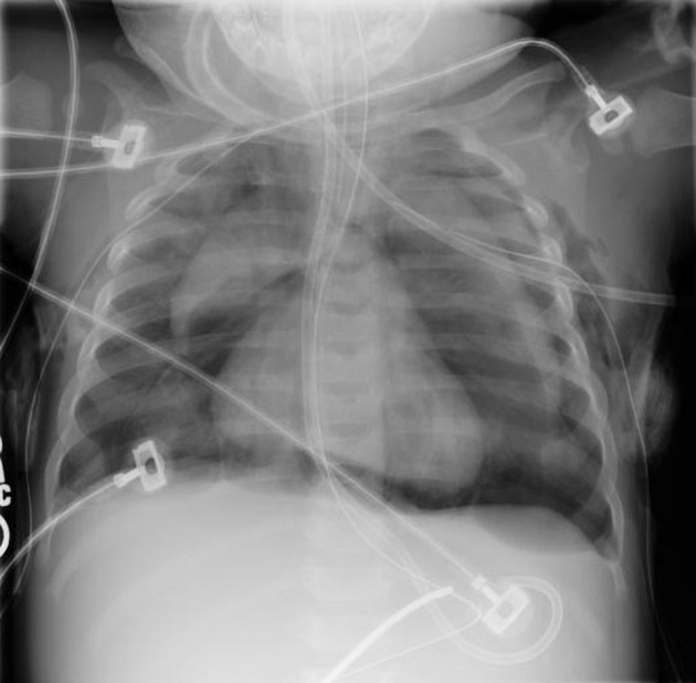
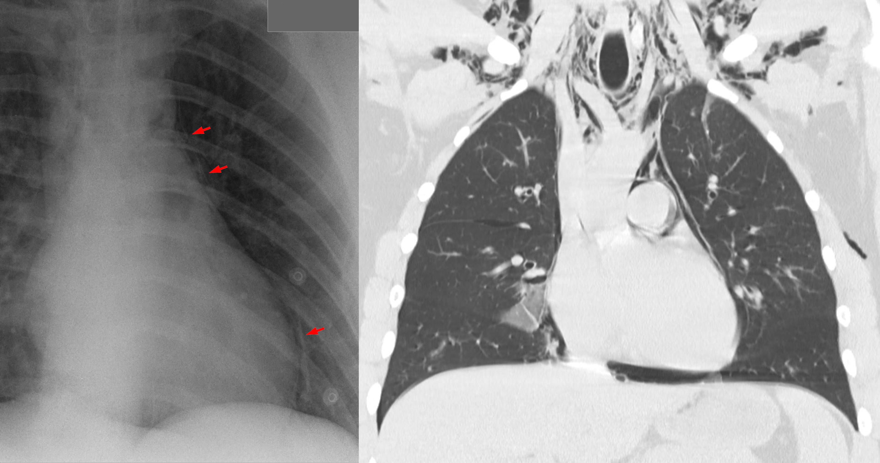
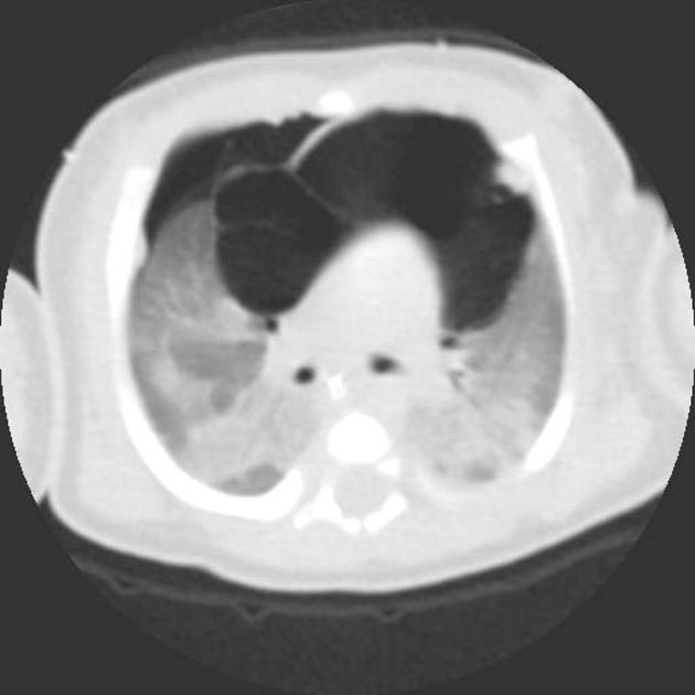
Small amounts of gas appear as linear or curvilinear lucencies outlining mediastinal contours such as:
- subcutaneous emphysema
- gas anterior to pericardium: pneumopericardium
- gas around pulmonary artery and main branches: ring around artery sign
- gas outlining major aortic branches: tubular artery sign
- gas outlining bronchial wall: double bronchial wall sign
- continuous diaphragm sign: due to gas trapped posterior to pericardium
- gas between parietal pleura and diaphragm: extrapleural sign
- gas in pulmonary ligament
- Naclerios V sign
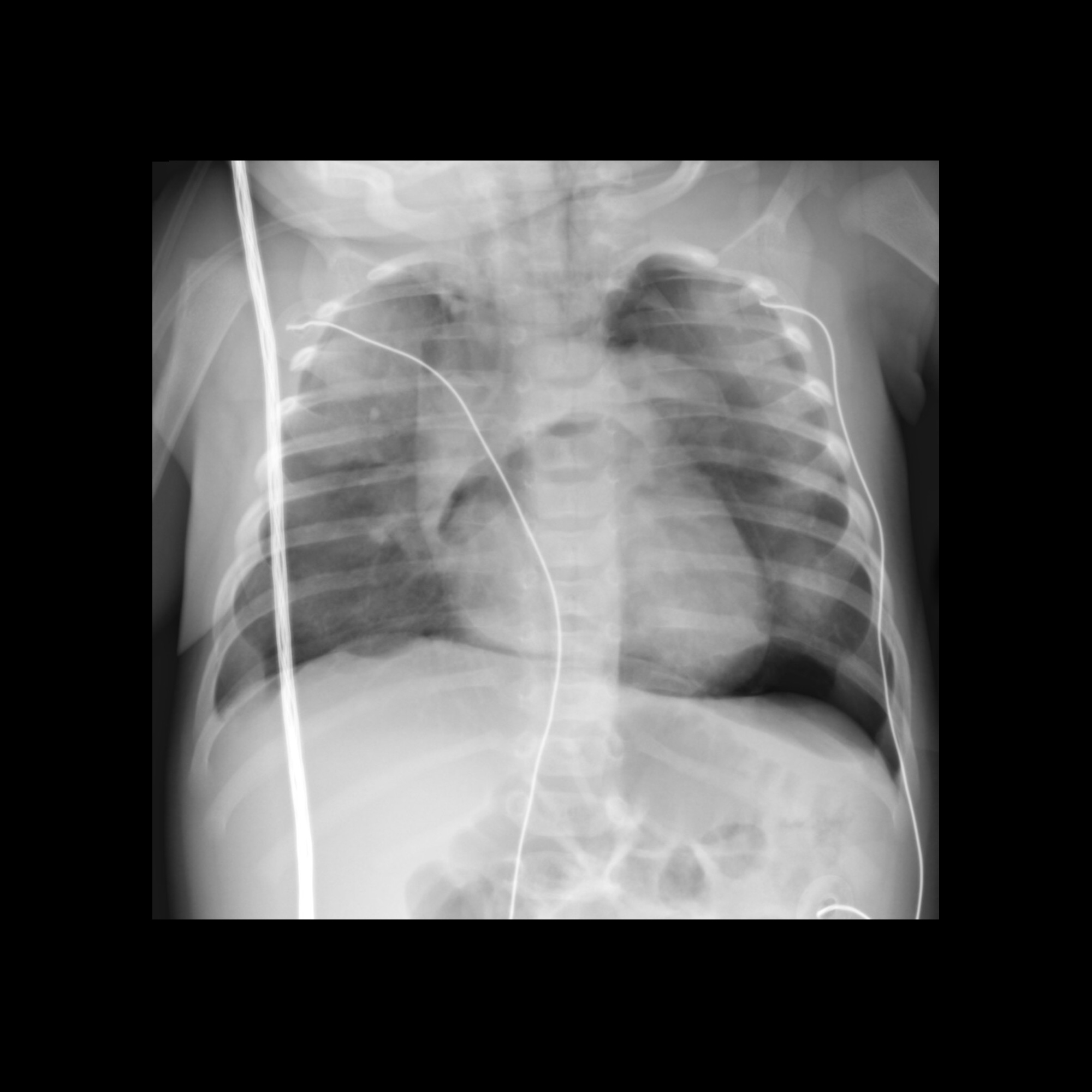
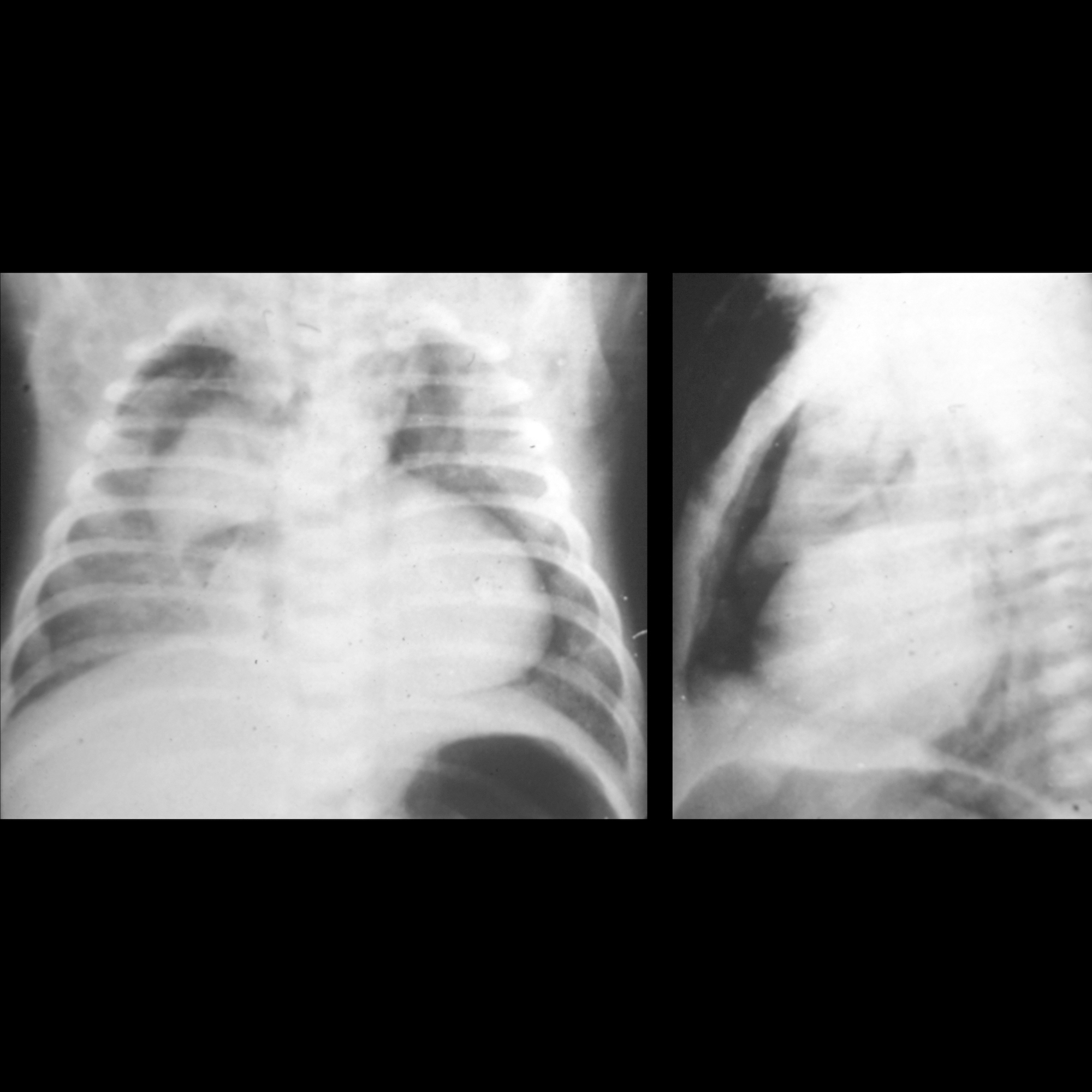
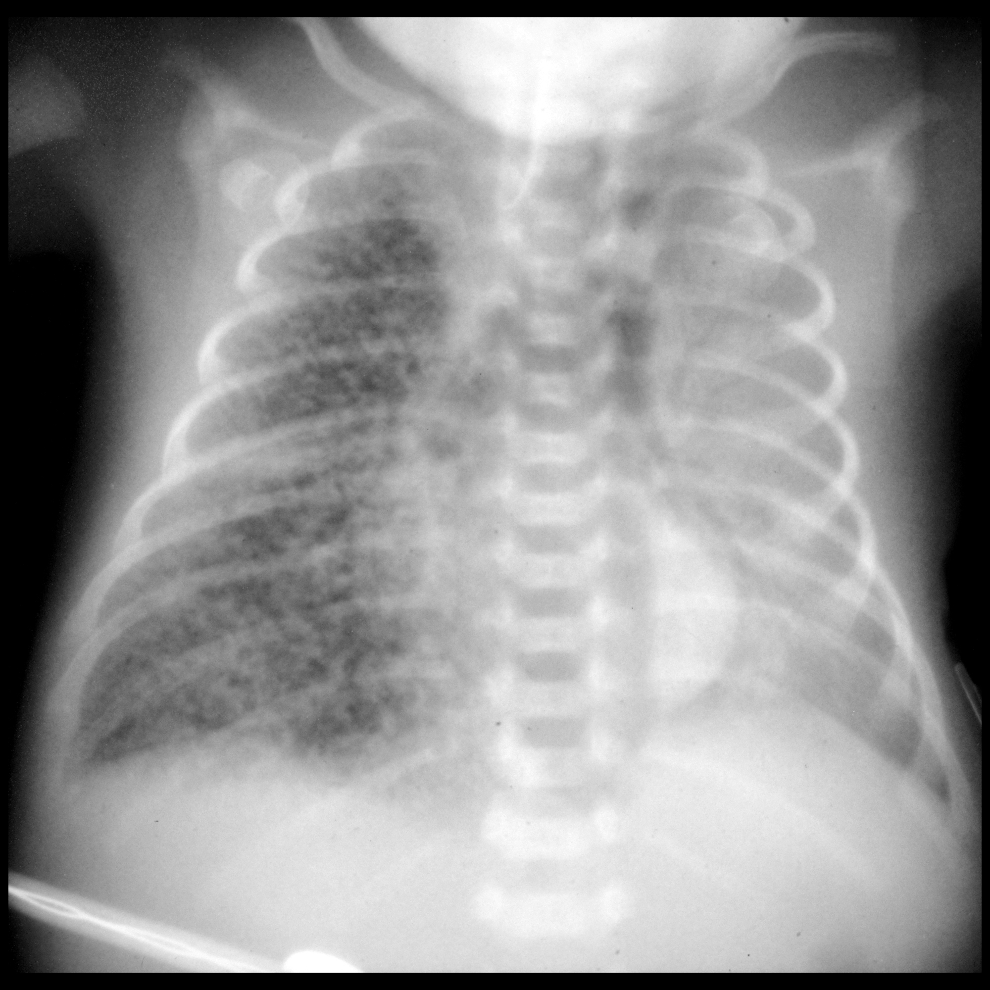
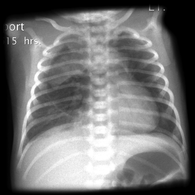
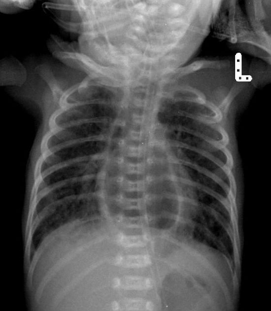
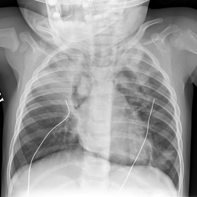
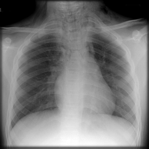
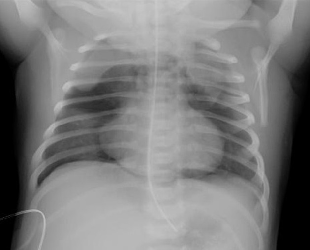
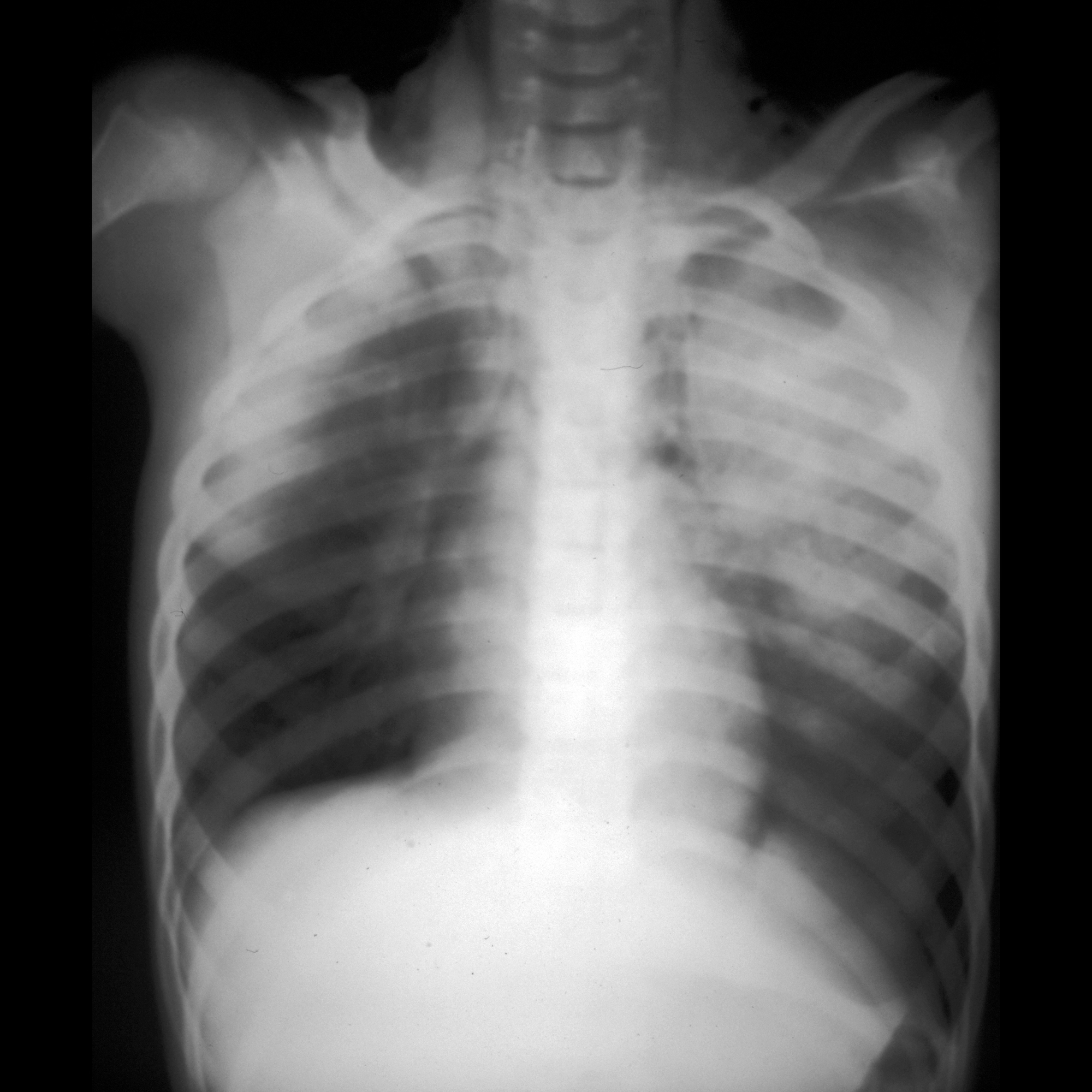
Pediatric pneumomediastinum may have slightly different appearances:
- elevated thymus: thymic wing sign
- gas crossing the superior mediastinum: haystack sign (the heart appears like a haystack in a Monet painting)
Ultrasound
Sonography is not a diagnostic modality of choice but is often used in the initial workup of undifferentiated trauma patients, or in differentiating causes of dyspnea or chest pain. Sonographic features which may appear in pneumomediastinum include :
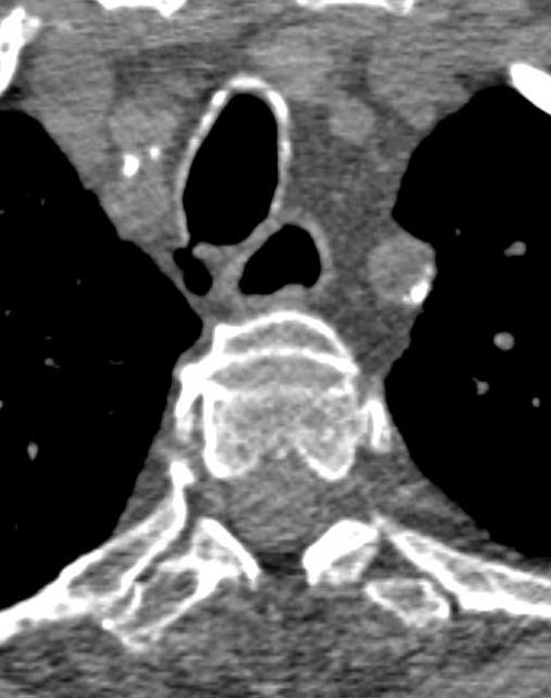
- cervical subcutaneous emphysema
- anterior cervical ultrasonography demonstrates hyperechoic foci with "dirty" posterior acoustic shadowing
- typically found anterior or lateral to the thyroid and medial to the internal jugular vein
- loss of the parasternal and apical views when performing transthoracic echocardiography
- with preservation of the subxiphoid window
- the air gap sign
- originally described using M-mode, describes systolic obscuration of cardiac structures as viewed from a parasternal long axis view
- cyclic appearance of a dense band of horizontal reverberation artifacts which were discontinuous in the far field
Differential diagnosis
Must be distinguished most importantly from:
- medial pneumothorax
- pneumopericardium
- pneumoperitoneum
- pneumoretroperitoneum
- Mach bands
- subcutaneous emphysema
For small gas collections on a CT scan, also consider :
Siehe auch:
- Pneumothorax
- Trachealdivertikel
- Pneumoperikard
- interstitielles Lungenemphysem
- Mach effect
- prävertebrales Weichteilemphysem nach Zahnbehandlung
- Macklin-Effekt bei stumpfem Thoraxtrauma
- Mediastinalemphysem beim Neugeborenen
- Mediastinalemphysem bei einem Kind
und weiter:
- Pneumoperitoneum
- Mediastinum
- Langerhanszell-Histiozytose der Lunge
- Spinnaker-Zeichen
- Weichteilemphysem
- Chronisch obstruktive Lungenerkrankung
- continuous diaphragm sign
- PLCH
- angel wing sign
- Boerhaave-Syndrom
- PIE
- Bronchialdivertikel
- subcarinale Bronchialdivertikel
- intraspinale Luft / Gas
- spontanes Pneumomediastinum
- pneumomediastinum from ingested fish bone
- blunt traumatic pneumomediastinum and subcutaneous emphysema
- perforated sigma diverticulitis causing mediastinal emphysema
- blunt tracheal rupture with extensive pneumomediastinum
- blunt traumatic pneumomediastinum outlining thymic remnants
- Postpartum pneumomediastinum
- Barotrauma der Lunge
- tracheobronchiale Verletzungen
- bronchial fracture (mnemonic)
- Pneumomediastinum/Pneumothorax - iatrogen
- pneumomediastinum as a complication of asthma
- Pneumomediastinum beim Neugeborenen
- pneumomediastinum in a child
- Mediastinalverbreiterung


 Assoziationen und Differentialdiagnosen zu Mediastinalemphysem: