renal vein anomalies
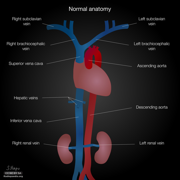
There are several variations in renal venous anatomy. Some of these are specific to the left renal vein.
Left renal vein anomalies are generally classified into four types:
- type I
- the ventral preaortic limb of the left renal vein is obliterated, but the dorsal retroaortic limb persists and joins the IVC in the normal position
- type II
- results from the obliteration of the ventral preaortic limb of the left renal vein and the remaining dorsal limb turns into a retroaortic left renal vein (RLRV)
- left renal vein lies at the level of L4 to L5 and joins the gonadal and ascending lumbar veins before joining the IVC
- type III
- is the circumaortic left renal vein or venous collar - due to the persistence of subsupracardial and intersupracardial anastomoses and the dorsal limb of the left renal vein.
- if all small retroaortic veins that empty into the IVC are considered, the incidence of a circumaortic left renal vein could be as high as 16%
- type IV
- the ventral preaortic limb of the left renal vein is obliterated, and the remaining dorsal limb becomes the RLRV
- other (non-classified): can involve either kidney
- supernumerary renal veins
- late venous confluence
See also
Siehe auch:
- retroaortaler Verlauf der linken Nierenvene
- Nussknacker-Syndrom
- developmental anomalies of the kidney and ureter
- circumaortic renal collar
und weiter:


 Assoziationen und Differentialdiagnosen zu renal vein anomalies:
retroaortaler
Verlauf der linken Nierenvene