Thymus
The thymus (plural: thymi) is a T-cell producing lymphoid organ in the anterior mediastinum that plays a role in the development of the immune system, particular the maturation of T-cells. It typically has a retrosternal location and hence may mimic retrosternal pathology.
Gross anatomy
It is relatively large in infancy (weighing 25 g at birth), grows considerably immediately after birth, and reaches a maximal weight in adolescence between 12 and 19 years (35 g). It gradually involutes with age (between 20 and 60 years) with progressive fatty replacement of the cellular components (15 g at 60 years of age). Fatty replacement starts at puberty and occurs more rapidly in males than females. There can be a wide variation in size between patients .
When sizable, it consists of two lateral lobes touching in the midline, situated partly in the thorax, partly in the neck. The two lobes are usually asymmetric in size. They are occasionally united, so as to form a single mass. Sometimes they are separated by an intermediate lobe.
The gland extends from as high as the lower border of the thyroid gland to the fourth costal cartilage downwards.
Relations
- anteriorly: sternum, origins of the inferior strap muscles
- posteriorly: pericardium, aortic arch, great vessels, left brachiocephalic vein, trachea
- laterally: pleura, pretracheal fascia
Arterial supply
Venous drainage
Lymphatic drainage
- parasternal, brachiocephalic and tracheobronchial lymph nodes
Innervation
- vagal fibers
- sympathetic fibers entering with blood vessels that are vasomotor
Histology
The thymus is of a pinkish-gray color, soft, and lobulated on its surfaces.
Development
Embryologically it is derived from the third pharyngeal pouch. The thymus is the first of the lymphoid organs to be formed. Considerable growth occurs immediately after birth in response to antigen stimulation and demand for mature T cells. Genetic factors also influence dependence upon thymus immunological function. After fibrofatty atrophy, the thymus can grow back at any time in life, particularly after periods of stress.
Variant anatomy
- variable location: ectopic and/or accessory thymic tissue may be located anywhere along the path of descent of the thymopharyngeal ducts, e.g. retrocaval, cervical, posterior mediastinal
- variable shape: e.g. unilobed, trilobed, X-shaped, inverted V-shaped, etc.
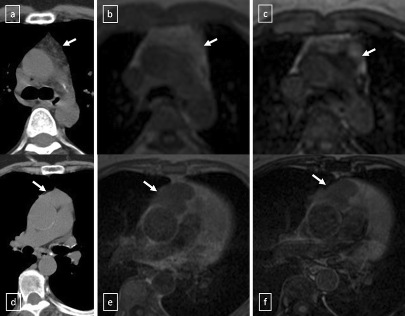
Radiographic features
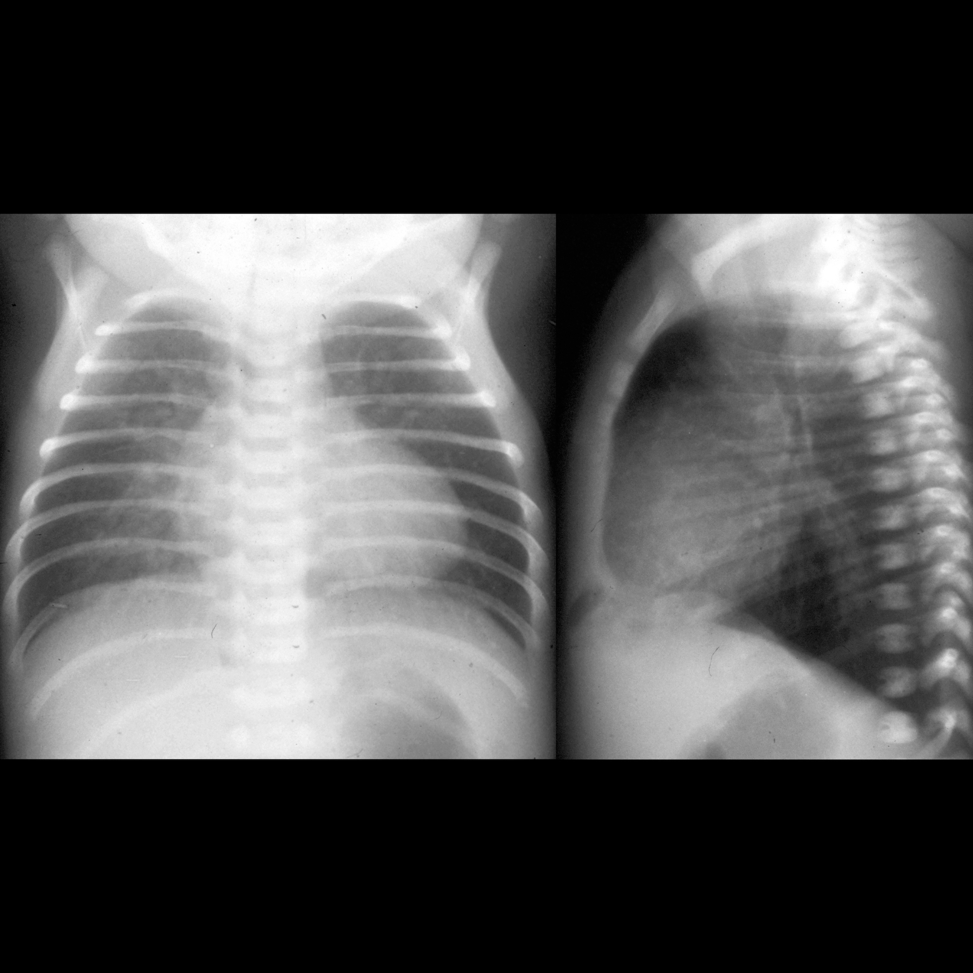
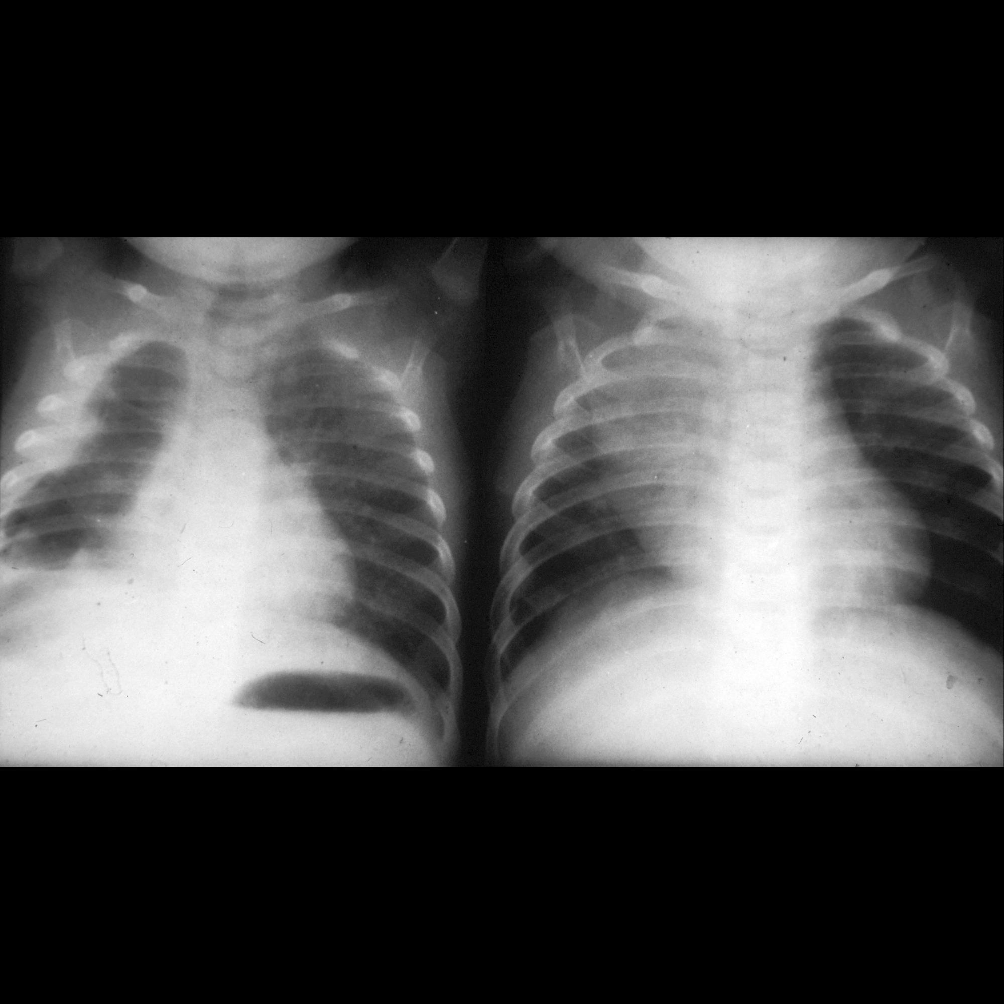
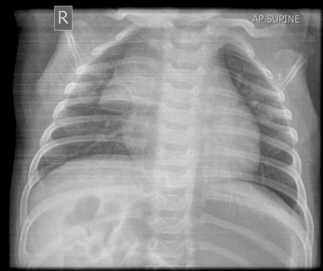
Plain radiograph
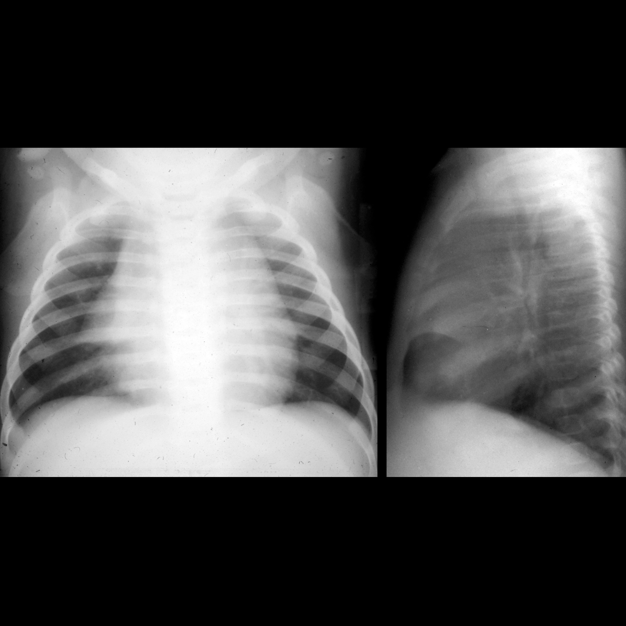
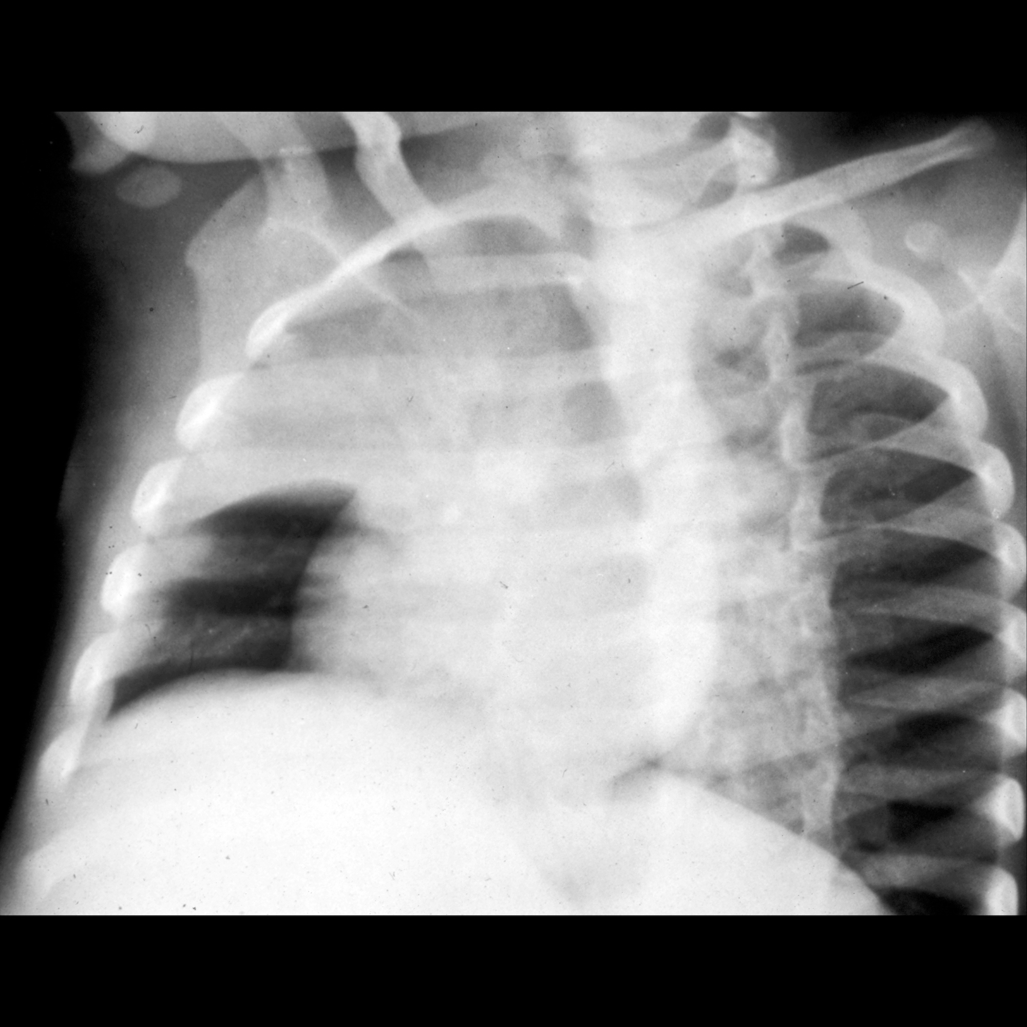
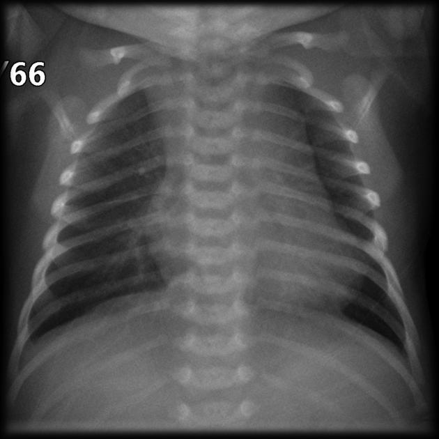
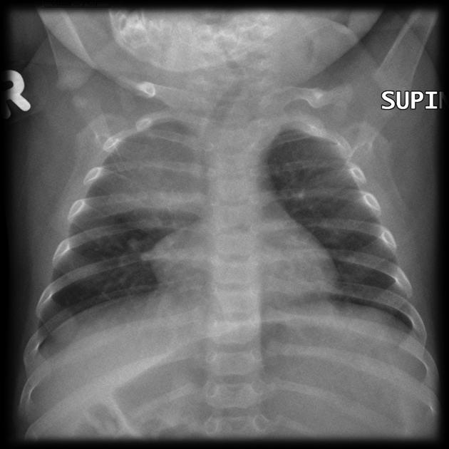
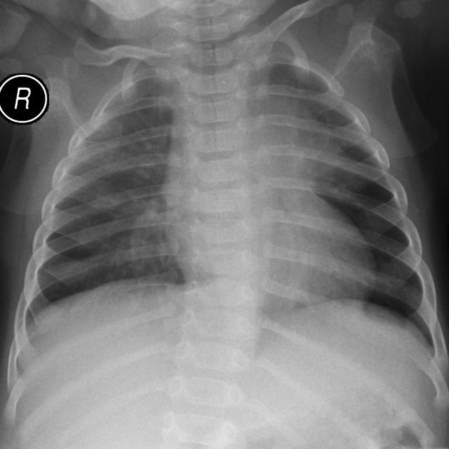
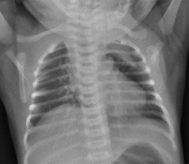
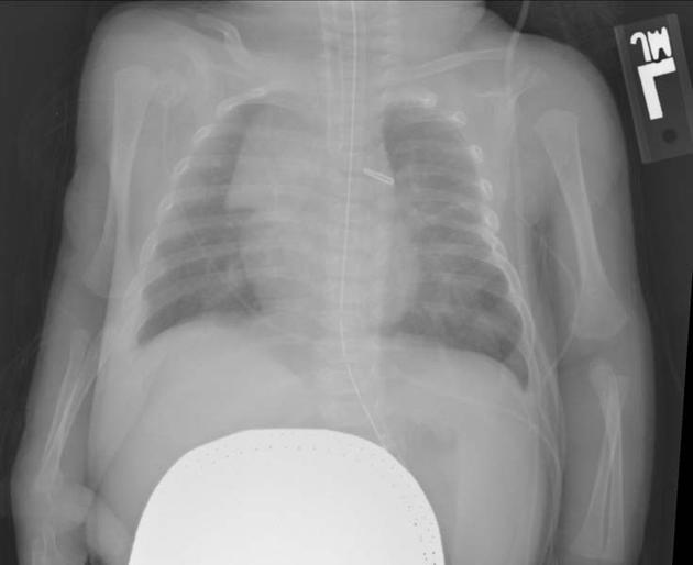
The thymus is seen as a triangular sail (thymic sail sign) frequently towards the right of the mediastinum. It has no mass effect on vascular structures or airway. The size can vary with inspiration.
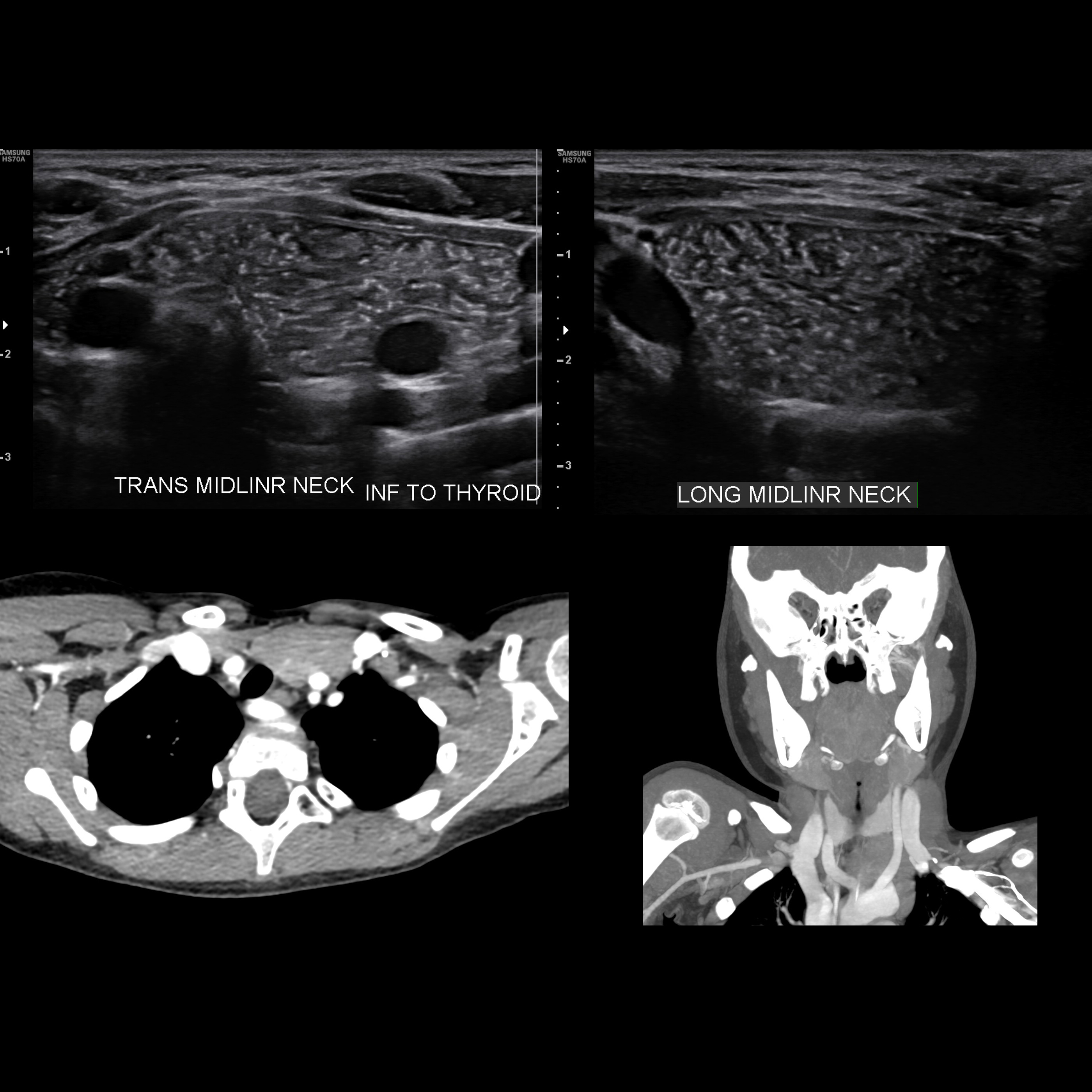
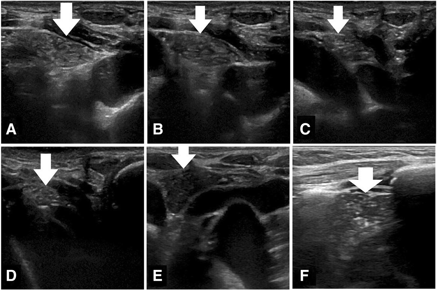
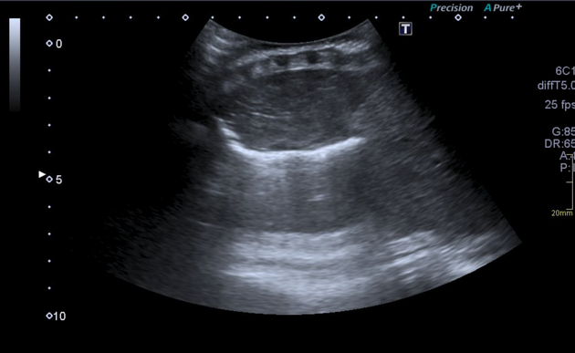
Ultrasound
typically relatively homogeneous background echogenicity similar to or slightly less than that of the liver and spleen, with scattered hyperechoic foci resembling a starry sky
shape can be affected/distorted by cardiac pulsations and respiratory motions as it is pliable and should not compress or displace adjacent structures
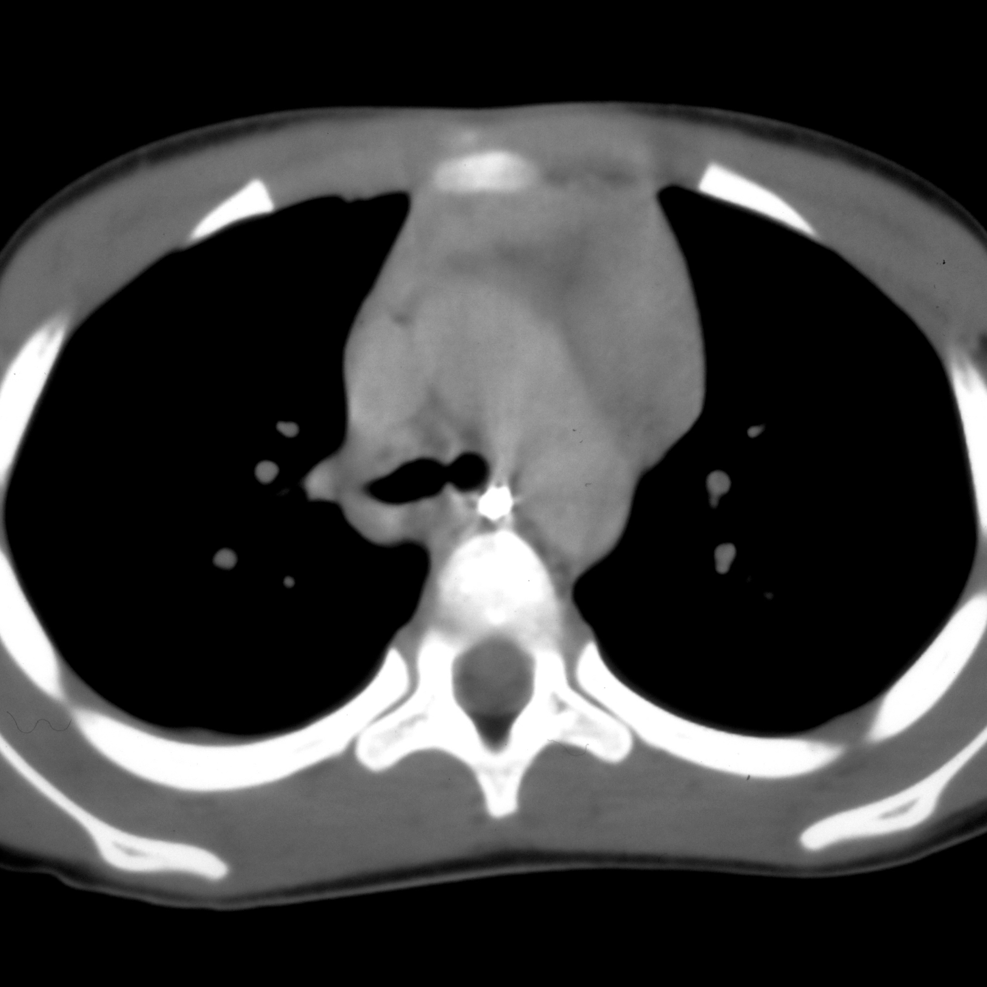
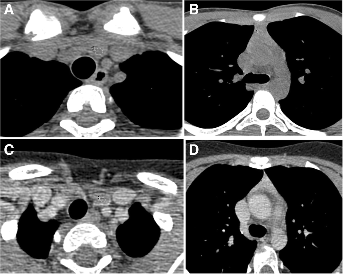
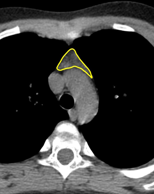
CT
- typically uniformly soft tissue density, approximately 80 HU and isoattenuating to surrounding muscle
- smooth outline with convex borders in childhood
- triangular in adulthood
- small blood vessels may be seen traversing it
MRI
- typically demonstrates chemical shift artefact between in and out of phase images
- differentiating normal from hyperplastic thymus can be difficult and guidelines for making this distinction and verifying the presence of normal thymus include :
- absence of rounded soft-tissue masses >7 mm
- absence of a convex contour of the thymus >19 years of age
- absence of soft-tissue lobulation
- absence of excessive thymic thickness (should be ≤1.3 cm when >20 years of age
- absence of a diagnosis associated with thymic enlargement or hyperplasia, e.g. Graves disease
History and etymology
"Thymus" ultimately derives from the Greek word for the plant "thyme" θύμος ("to offer/sacrifice"), presumably because the plant was burnt on altars. Galen thought the thymus gland looked like a "warty excrescence" and resembled a bundle of the plant .
The first good description of the thymus gland was recorded by Berengarius in 1524.
Related pathology
- thymic rebound
- thymic epithelial tumors
- primary tumors of the thymus
- thymic hyperplasia
- DiGeorge syndrome
- the normal thymus can mimic several pathologies:
- retrosternal hematoma
- spinnaker sign in pneumomediastinum
- aortic injury
Siehe auch:
- Schilddrüse
- Thymushyperplasie
- Mikrodeletionsyndrom 22q11
- thymic epithelial tumours
- vorderes Mediastinum
- normal thymic uptake of I-131
- ektopes Thymusgewebe
- Reboundhyperplasie des Thymus
und weiter:
- Mediastinum
- Tumoren des vorderen oberen Mediastinums
- Spinnaker-Zeichen
- mediastinal lymphoma
- Thymuslipom
- angel wing sign
- primäre Thymusneoplasien
- anterior junction line
- thymic carcinoid tumour
- Normale Herzkonfiguration im Röntgen-Thorax
- Teratom des Thymus
- Thorax Onlinekurs
- wave sign
- wavy thymus
- residual thymic tissue
- thymus - right sided
- Mediastinalverbreiterung


 Assoziationen und Differentialdiagnosen zu Thymus: