tumors of the paranasal sinuses

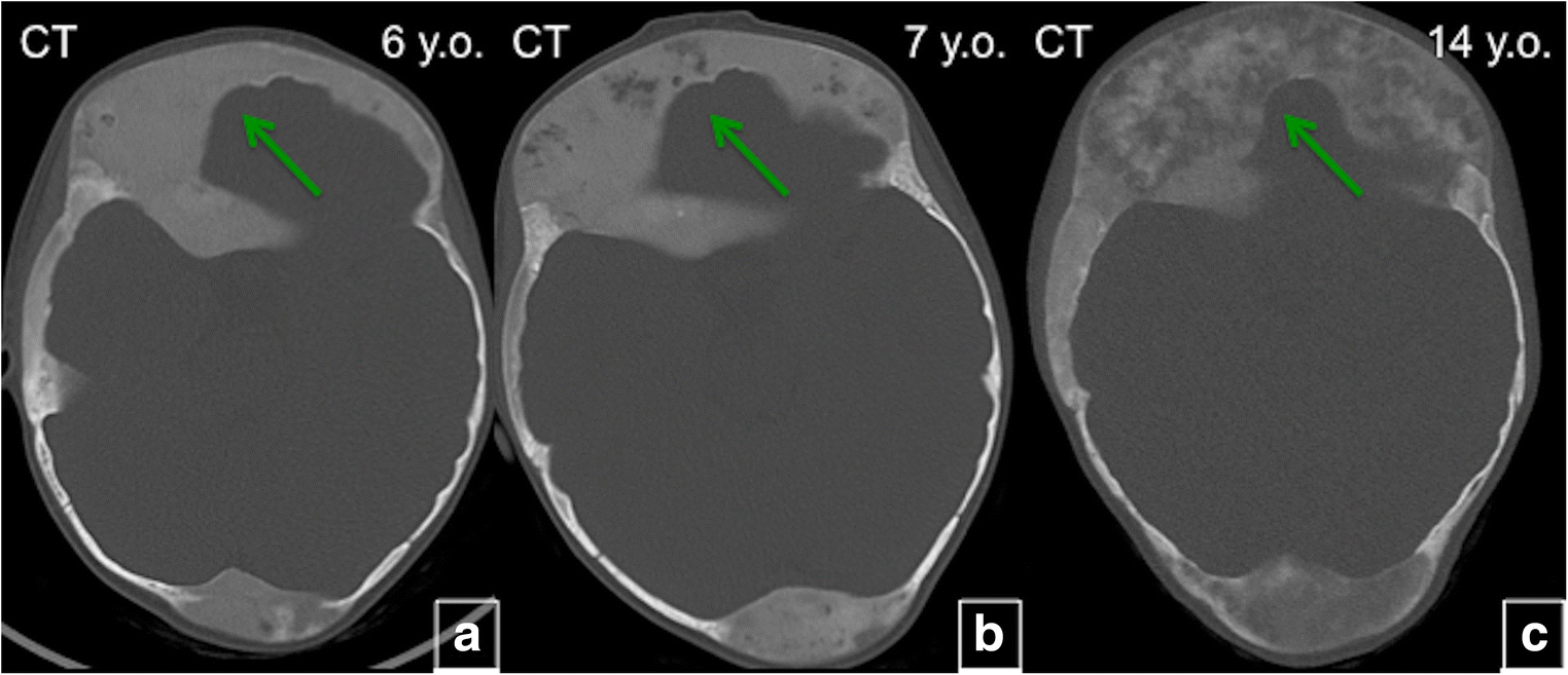
Giant cell
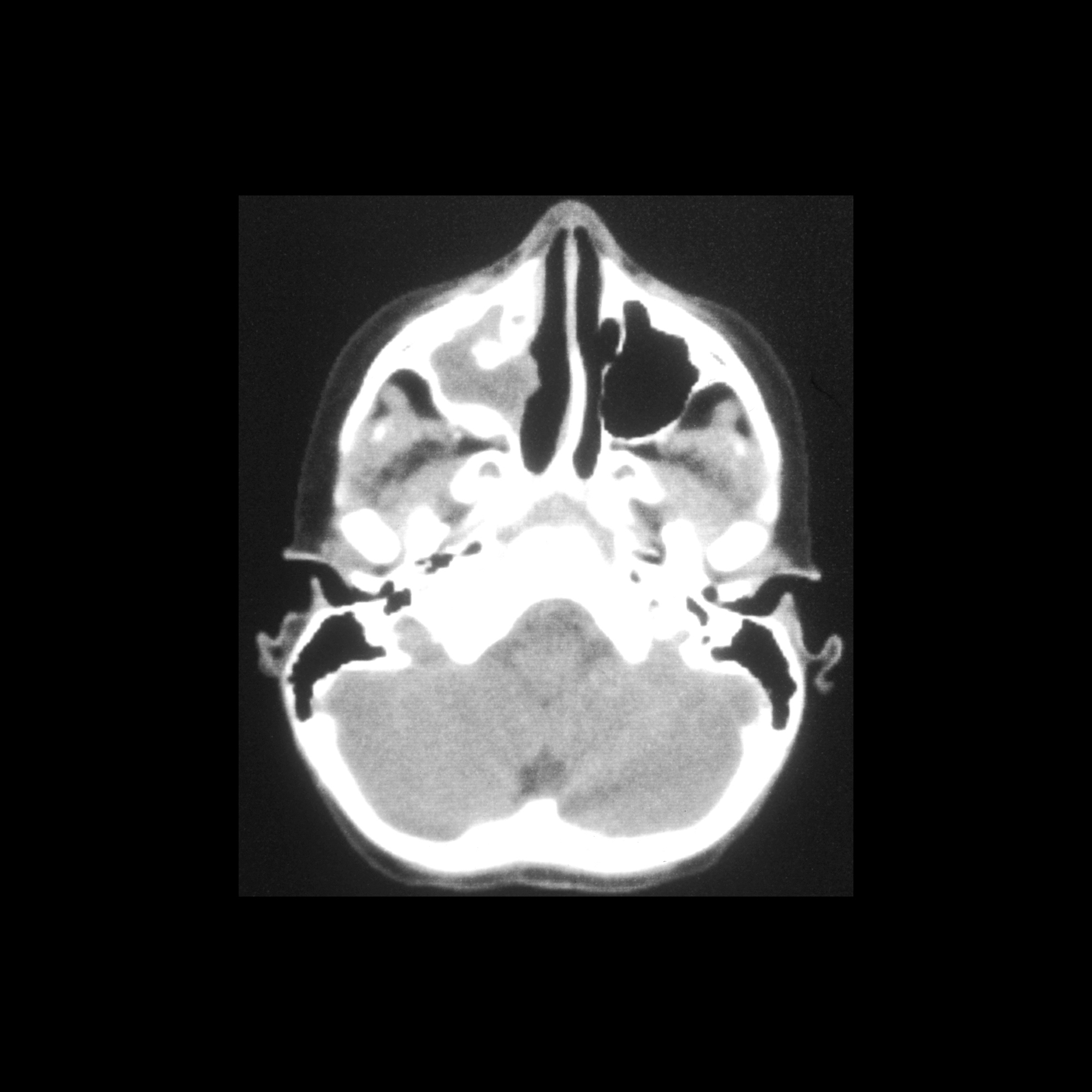
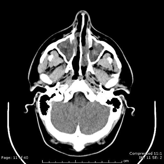
reparative granuloma of ethmoid sinus: A rare case report. Axial CT and MR (1 ABCD) images showing fluid fluid level in right half of ethmoidal sinus with proptosis of right eye.

Giant cell
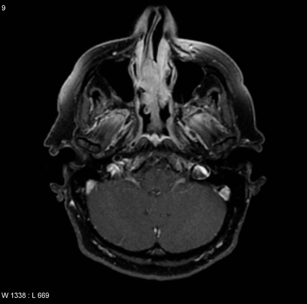
reparative granuloma of ethmoid sinus: A rare case report. T1 W axial images showing fluid fluid level.

Giant cell
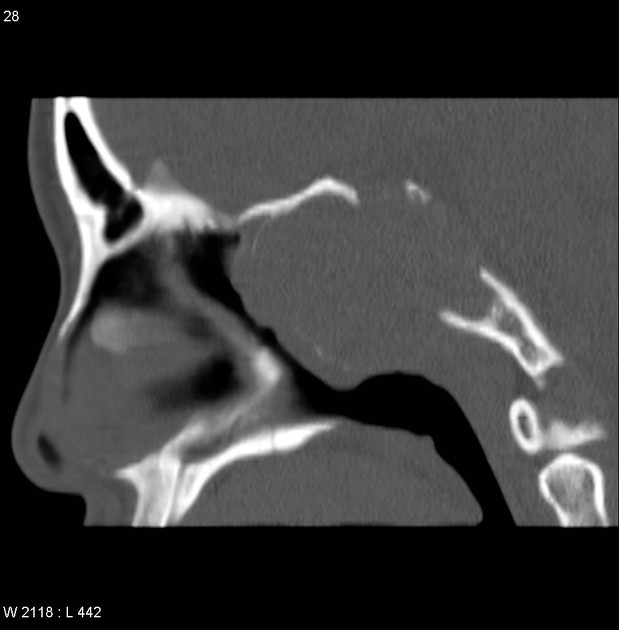
reparative granuloma of ethmoid sinus: A rare case report. Sagittal CT and MRI showing fluid fluid level.

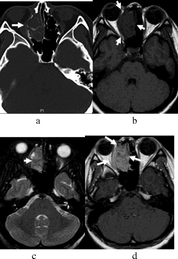
Sinonasal
myxoma – an unusual cause of a maxilary mass in a child. CT on bone (WL 600/WW 3000 - E-H) and soft tissue (WL 35/WW 280 - A-D) windows, 2.0 mm slice thickness, with axial, coronal and sagittal reformats.

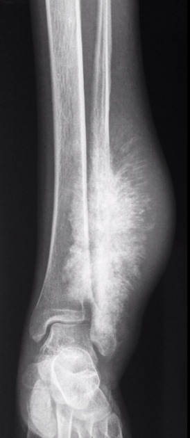
Sinonasal
myxoma – an unusual cause of a maxilary mass in a child. Axial, coronal and sagittal MRI, 1.5 Tesla, 5 mm slice thickness, T2 weighted (TR=5580ms TE=100ms), T1 weighted (TR=431ms TE=9.5ms) and diffusion weighted imaging (DWI and ADC map).
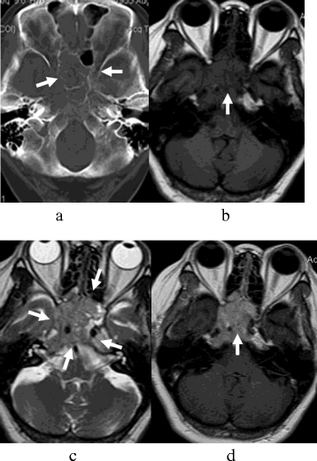
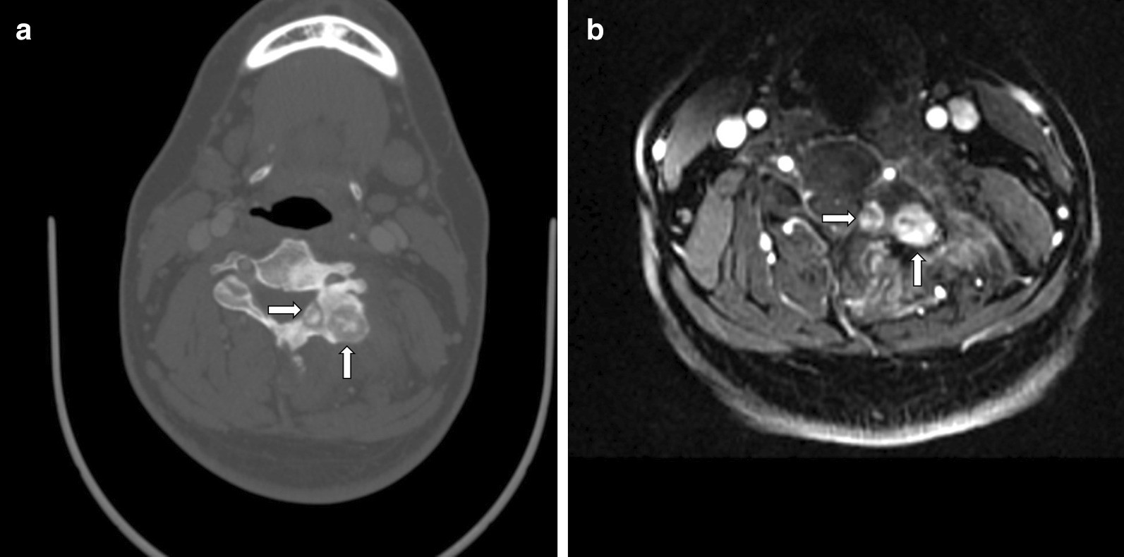
The CT and
MRI observations of small cell neuroendocrine carcinoma in paranasal sinuses. SNEC of paranasal sinuses in a 41-year-old man (a-d). The lesion was symmetrical, and the size was about 5.8 cm × 5.7 cm × 4.3 cm. (a) CT image showed worm-eaten bone destruction in sphenoid sinus, anterior cranial fossa, and orbital apex; however, bone contours still could be seen. (b) T1-weighted MR image demonstrated isointensity. (c) T2-weighted MR image demonstrated isointense together with a ‘pigeon’ pattern. (d) Contrast-enhanced T1-weighted MR image demonstrated a moderate heterogeneous enhancement mass, which showed involvement of the pharyngonasal cavity, orbital apex, pterygopalatine fossa, sella, cavernous sinus, internal carotid canal, and jugular foramen.
The CT and
MRI observations of small cell neuroendocrine carcinoma in paranasal sinuses. SNEC of paranasal sinuses in a 53-year-old man (a-d). The tumor size was about 4.3 cm × 4.1 cm × 3.1 cm. (a) CT image showed worm-eaten bone destruction in the right ethmoidal sinus and fossa orbitalis; however, bone contours still could be seen. (b) T1-weighted MR image demonstrated isointensity. (c) T2-weighted MR image demonstrated isointense mixture. (d) Contrast-enhanced T1-weighted MR image demonstrated a mild heterogeneous enhancement mass, which showed involvement of the pharyngonasal cavity and fossa orbitalis.
Tumoren der Nasennebenhöhlen
tumors of the paranasal sinuses
Siehe auch:
- Fibröse Dysplasie
- Osteosarkom
- Osteom NNH
- Osteoblastom
- Sinunasale Aspergillose
- Mukozele der Nasennebenhöhlen
- Invertiertes Papillom
- Nasopharynxkarzinom
- Plattenepithelkarzinom der Nasennebenhöhlen
- Plattenepithelkarzinom des Sinus maxillaris
- Non-Hodgkin-Lymphom des Sinus maxillaris
- Adenokarzinom der Nasennebenhöhlen
- solitary fibrous tumour in the paranasal sinuses
- Tumoren der Kieferhöhle
- Myxom der Nasennebenhöhlen
- Polyposis der Nasennebenhöhlen
- Tumoren der Nasenhaupthöhle
- kleinzelliges neuroendokrines Karzinom der Nasennebenhöhlen
und weiter:


 Assoziationen und Differentialdiagnosen zu Tumoren der Nasennebenhöhlen: