Left ventricular hypertrophy
Left ventricular hypertrophy (LVH) is present when the left ventricular mass is increased. It is a common condition, typically due to systemic hypertension, and it increases with age, obesity and severity of hypertension.
Epidemiology
Studies have demonstrated a prevalence on echocardiography of 36-41% in hypertensive patients .
Clinical presentation
When mild, left ventricular hypertrophy is usually asymptomatic. As it worsens, symptoms of congestive heart failure gradually arise.
Pathology
Left ventricular hypertrophy may result from either increased pressure or volume afterload on the heart. Much more rarely it may result from genetic conditions. The relationship between obesity and left ventricular hypertrophy is complex.
Etiology
A large number of conditions can cause left ventricular hypertrophy.
- hypertension: pressure overload
- systolic BP >140 mmHg
- diastolic BP >90 mmHg
- valvular heart disease
- aortic stenosis: pressure overload
- aortic regurgitation: volume overload
- mitral regurgitation: volume overload
- obesity
- genetic diseases
- severe physical exercise
Radiographic features
Echocardiography, cardiac MRI and cardiac CT are the main modalities for evaluating for left ventricular hypertrophy.
Robust reference data exists for quantifying left ventricular size with echocardiography, using guidelines from the American Society of Echocardiography and the European Association of Cardiovascular Imaging. Similar validated data does not (yet) exist for cardiac MRI/CT and therefore most use the echo data for measurements on MRI and CT.
The primary parameters employed to assess the degree of left ventricular hypertrophy are:
- wall thickness
- left ventricular mass (LVM)
- left ventricular geometry
Echocardiography
Echocardiography (echo) is the key tool for assessing left ventricular hypertrophy. This is due to its universal availability, absence of ionizing radiation and superb temporal resolution, the latter surpassing all other contemporaneous techniques; modern M-mode echo can achieve 1 ms resolution. Like any ultrasound-based technique, echocardiography is more dependent on the operator than CT/MRI and is more affected by patient factors. In general a transthoracic approach suffices in the majority of patients, in a small minority, e.g. emphysema, a transesophageal technique may be necessary.
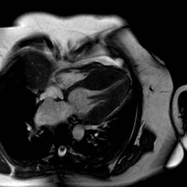
Cardiac MRI
Cardiac MRI (CMR) is the current gold standard for evaluation of left ventricular hypertrophy, with the ability to measure multiple other myocardial parameters simultaneously. Its spatial resolution is poorer than echo/cardiac CT, takes longer to perform than either echo or CT, and has relative limitations in those with implanted cardiac devices (e.g. mechanical valves, pacemakers, etc.).
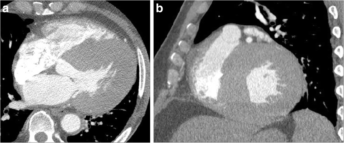
Cardiac CT
Cardiac CT is currently the least favored technique for assessment of left ventricular hypertrophy due to the relatively high radiation dose. It is a fast technique, with high spatial resolution, although its temporal resolution is poor.

