conventional intramedullary chondrosarcoma
Conventional chondrosarcoma also known as central chondrosarcoma is the most common subtype of chondrosarcoma and may be low, intermediate or high grade (see chondrosarcoma grading).
Epidemiology
They typically occur in the 4 and 5decades with a slight male predominance 1.5-2:1.
Clinical presentation
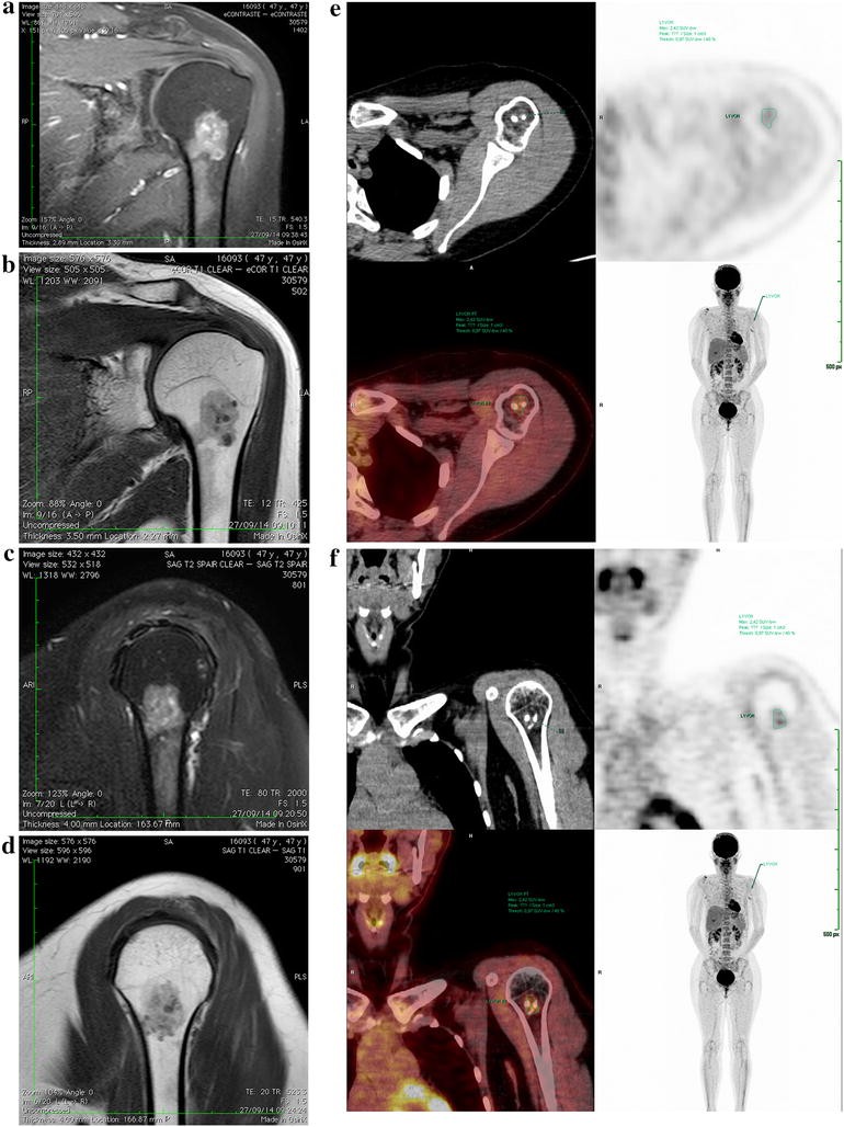
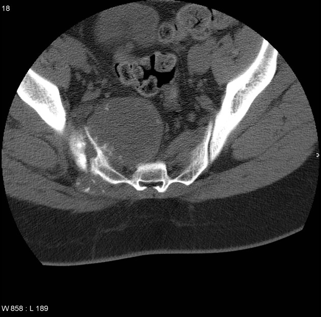
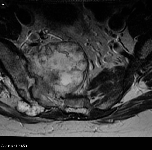
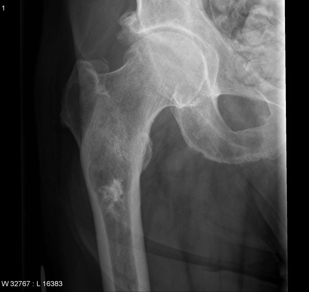
At diagnosis it is typically a large mass, usually over 4 cm in diameter. When arising in long bones (most common location) it typically involves more than 50% of the length of the shaft.
Typically chondrosarcomas present with:
- pain: present in 95% of cases, often long standing and worse at night
- palpable mass: present in 28-82% of cases
- pathological fracture: present in 3-17% of cases
Pathology
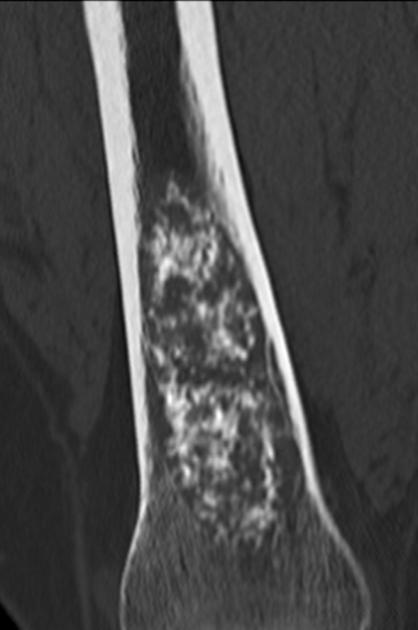
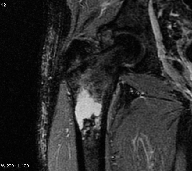
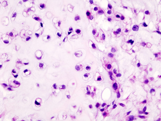
Histologically, the tumor grows as multiple hyaline cartilage nodules with central high water content and peripheral endochondral ossification. This accounts for not only the high T2 MRI signal but also for rings and arcs calcification and popcorn calcification on CT and plain film.
Location
Radiographic features
For imaging findings please refer to the article on chondrosarcoma.
Differential diagnosis
- enchondroma
- low-grade conventional chondrosarcomas can be difficult to distinguish from an enchondroma, as both grow in a nodular pattern and result in scalloping of the inner surface of the cortex
- scalloping of greater than two-thirds of the cortical thickness, cortical breach and soft tissue mass beyond the confines of the bone are useful distinguishing features
- see article: enchondroma vs. chondrosarcoma
Siehe auch:
- Enchondrom
- Chondrosarkom
- popkornartige Verkalkungen
- Unterscheidung Enchondrom Chondrosarkom
- chondrosarcoma grading
- ring- und bogenförmige Verkalkungen
und weiter:


 Assoziationen und Differentialdiagnosen zu conventional intramedullary chondrosarcoma: